Pathophysiology of Shock

In a normal human physiologic state, there is a homeostasis that exists in cells that includes good stores of energy (e.g. ATP) and energy sources (e.g. glucose), an adequate supply of oxygen, and a pH , fluid, and electrolyte balance that optimizes cellular function. About 20% of the energy expended in the body at rest, that is 20% of the basal metabolic rate (BMR), is used to maintain Na+ and K+ gradients via the Na+/K+ ATPase pump. In fact, the brain and kidneys use 50 – 70% of their BMR on this function.  If any of the conditions above are disrupted, say by lack of blood flow to cells of the body, cellular dysfunction occurs.  If disruption persists for a long enough period of time functions, such as the Na+/K+ ATPase pump on the cell membrane, begin to fail and Na+ and K+ (along with fluid) follow diffusion gradients and begin to equilibrate between the intracellular space and interstitial space.  Homeostasis is lost, enzymes fail to function properly, and cellular death occurs.  The goal, then, is to recognize and treat shock when it first manifests.  This will decrease morbidity and mortality in your patients.

So, what is shock?  Clearly stated, shock is defined as a decrease in circulation of oxygen and/or nutrients to tissues to the point that those tissues malfunction, become damaged, and ultimately, in severe cases, are destroyed.  In order to avoid shock, then, one must have adequate blood flow to the tissues, and that blood has to be carrying adequate oxygen and nutrients.  In resuscitation of any critically ill or injured patient, the physician must address the ABC’s: i.e. Airway, Breathing, and Circulation.  If the patient cannot maintain a clear airway, one must be provided, often by placement of an endotracheal tube. Breathing, if inadequate, must be assisted. If the patient is hypoxic (has low oxygen in the blood), supplemental oxygen is administered as well. The third component of this triad, circulation, is central to the pathophysiology and treatment of shock. 

  There are three factors that determine blood flow to tissues: blood volume, “pump function” (cardiac function), and vascular capacitance and resistance.  Low blood volume, due to hemorrhage or dehydration, results in a decrease cardiac output because there is a decrease in stroke volume. When hydrostatic pressure is reduced to the point that perfusion to the body is compromised, we call this hypovolemic shock. Cardiogenic shock is poor tissue perfusion as a result of the heart not functioning well. This decrease in cardiac output can be the result of decreased stroke volume (e.g. from a heart attack and impaired contractility of the heart muscle), a low heart rate (e.g. from an unfavorable drug effect), or valve dysfunction, where forward flow of blood is impeded.  Vasogenic shock refers to decreased perfusion to the core of the body because of some problem with vascular tone. Under normal circumstances there is vascular tone, or slight vasoconstriction, to the extremities. This is because they do not need a lot of blood flow when at rest. When we exercise, additional flow is needed in the extremities and thus we increase our cardiac output, by increasing our heart rate, to accommodate this need. There are conditions, though, that occur when there is abnormal dilatation of the blood vessels (e.g. toxic effect of drugs), where perfusion to the core is compromised because of dilatation of arteries to the periphery. In the case of bad infections, sepsis, there can also be leakage of fluid from capillaries because of effects of certain conditions created. In this instance, intravascular proteins can be leaked into the interstitial space and fluid will be drawn out of the vascular space (since there is a loss of intravascular oncotic pressure). These vascular problems result in vasogenic shock.

There is one more type of shock that does not fit neatly into any of the above categories and that is something commonly referred to as mechanical shock. In this instance, there is impedance of blood flow as a result of something physically impairing blood flow. This can be the result of a collapse of the lung causing a tension pneumothorax, pulmonary emboli, cardiac tamponade, or some kind of mass or tumor compressing a major vessel from the outside and impairing flow. Again, when any of these conditions exist, we refer to this kind of shock as, mechanical shock. 

The human body has some ability to compensate for adverse conditions leading to shock, e.g. heart rate can increase to improve cardiac output, blood vessels to the extremities can constrict to shunt blood preferentially to important organs (heart, lungs, and brain), and the nervous system can give a sense of thirst and distress.  Compensatory mechanisms, however, can quickly become overwhelmed, and a state of non-compensatory shock sets in.  If conditions do not improve promptly, terminal shock ensues, organs fail, and the victim dies.  These are the 3 stages of shock: 1. Compensatory, 2. Non-compensatory, and 3. Terminal.  Compensatory shock can be subtle.  The patient may be mildly anxious, diaphoretic (sweaty), and tachycardic (defined as a heart rate greater than 100 beats per minute in a normal adult).  The blood pressure will be normal or near normal (systolic pressure about 110 to 130 mmHg for normal adult) and the patient will be mentating normally.  During compensatory shock, the body is releasing endogenous catecholamines (e.g. epinephrine from the adrenal glands) in an effort to increase cardiac output and constrict blood vessels leading to the skin and extremities and shunt blood preferentially to the abdomen, chest, and brain.  In addition, the kidneys are attempting to retain as much fluid as possible, so urine output decreases.  As conditions worsen, compensatory mechanisms begin to fail.  Catecholamine stores are depleted, renal function fails because of poor perfusion, and the patient’s blood pressure drops further.  If the cause of shock is not targeted soon and treated, the patient will progress to terminal shock and ultimately die.  Terminal shock is a condition in which cellular function is actively failing because of poor perfusion to the tissues.

The treatment of shock is straightforward: identify that the patient is in shock, find the cause of shock, and treat the underlying condition of causing shock.  After stabilization of the patient’s condition, provide supportive care to sustain a more normal physiologic state.  With this fund of new knowledge, let’s take a look at our original case scenario that started this chapter and see if we can save his life!

Capillary Physiology

The cells of the body require a continuous supply of oxygen and nutrients to survive.  They also require a process by which wastes can be carried away.  It is the role of the circulatory system to carry out these vital functions, and it is at the level of the capillaries that this process takes place.  Capillaries do not exchange directly with the intracellular compartment, but instead with an interstitial space that lies between the capillary and the cell membrane.


This interstitial space is a milieu of nutrients and wastes that are continually being exchanged by the intracellular and intravascular forces acting upon them.  Capillary circulation reaches every living cell in the body, via the interstitial fluid, and allows the exchange of nutrients and wastes.  Though there are different types of capillaries, which allow different amounts of exchange with the interstitial space, the general principles of diffusion remain the same and will be discussed in general terms.  Capillaries are thin walled and allow small particles and water to flow freely from the intravascular space to the interstitial space through tiny slits following diffusion gradients.  Large particles (e.g.  blood cells, proteins, and other large masses) are trapped within the vascular space.  These “trapped particles” create an osmotic gradient (oncotic pressure) that draws fluid back into the vascular space.  That is to say, oncotic pressure counteracts the hydrostatic pressure that drives fluid out of the vascular space.  There are four factors then, under normal circumstances, which determine the diffusion of water and other particles across a capillary wall:

  1. Particle Size
  2. Hydrostatic Pressures
  3. Oncotic Pressures
  4. Concentration Gradients

Under normal circumstances, fluid, electrolytes, oxygen and other nutrients flow out of the proximal portion of a length of capillary, and into the interstitial milieu that exists surrounding all cells (Figure 16).  As blood travels further through this length of capillary, hydrostatic pressure decreases to a point at which it equals oncotic pressure, and there is equilibrium of exchange to the point that only diffusion gradients affect the transport of particles across the capillary wall.  At the end, or distal portion, of a length of capillary, oncotic pressure is greater than hydrostatic pressure, and fluid is drawn back into the intravascular space (Figure 16).

EquilibriumBetweenIntravascularAndInterstitial-figure16

Figure 16
A normal state of equilibrium between the intravascular (filled with large particles, such as proteins, that creates an “oncotic” pressure) and interstitial spaces.

When hydrostatic pressures are high, there will be more fluid transported to the interstitial space (Figure 17).  When hydrostatic pressures are low, there will be more fluid transported into the intravascular space (Figure 18).  When oncotic pressures are low (e.g. in cases in which the liver does not produce enough albumin, such as in patients with liver failure), even with normal hydrostatic pressures, there will tend to be more fluid transferred to the interstitial space (Figure 19).  These principles are important because normal intracellular function depends on a very narrow range of fluid balance, electrolyte concentrations, and pH, and requires a continuous supply of oxygen and nutrients.  If any of these are disrupted, it will lead to cellular dysfunction and ultimately to cell death as conditions worsen.

EquilibriumBetweenIntravascularAndInterstitial-figure17

Figure 17
Elevated hydrostatic pressures in the capillary moves the point of equilibrium of Hydrostatic and oncotic pressures to the right (toward the venous end of the capillary) and allows less time for fluid and wastes to re-enter the circulatory
system. This leads to fluid retention in the tissues (edema).

EquilibriumBetweenIntravascularAndInterstitial-figure18

Figure 18
In a state of low hydrostatic pressure, the intravascular oncotic pressure is relatively higher thus the equilibrium between these opposing forces is moved to the left. This results in greater movement of fluid from the interstitial space to the intravascular space.

EquilibriumBetweenIntravascularAndInterstitial-figure19

Figure 19
When there are less intravascular osmotically active particles (less protein), this results in a state of low oncotic pressure. The hydrostatic and oncotic pressures equal each other closer to the venous end of the capillary. This results in a greater amount of time for fluid to escape from the vascular space and thus leads to tissue edema, much like high hydrostatic pressure does. Note: We don’t discuss a state of high oncotic pressure because it would be a very rare condition in which that would occur. If it did happen, though, what would the illustration look like?

Concentration gradients (Figure 20) exist between the interstitial space and the intracellular space because of complex “pump systems” that exist on the cell membrane of every human cell.  One of the most important pump systems is the Na+/K+ ATPase pump.  This pump creates an osmotic gradient between the high concentration of Na+ outside the cell, and the high concentration of K+ inside the cell.  It takes a lot of energy for cells to maintain these concentration gradients, and these gradients are necessary to optimize intracellular enzymatic function and to facilitate transport of certain interstitial particles into the cell.  There is an equilibrium between the intravascular space and the interstitial space such that the concentrations of most substances are essentially equal to each other, except for those particles that cannot escape the intravascular space because of their large size (e.g. blood cells, protein molecules).  We will discuss this topic again in the renal chapter.

ConcentrationGradients-figure20

Figure 20
The intravascular (plasma) space and the interstitial space share similar concentrations of ions, with sodium (Na+) being the primary cation and Chloride (Cl-) and bicarb (HCO3-) being the predominant anions (electrolytes measured in meq/L). The intracellular space has a much different
composition of ions because the cell membrane tightly controls the movement of charged particles into and out of the cell. The primary intracellular cation is potassium (K+) and the predominant intracellular anions are organic phosphates and proteins.

The Perfect Medical School Application

Walking up steps take 2

You’re putting your medical school application together, going down the list checking off the various items, and you realize you’ve done pretty well:  You’ve maintained a decent GPA and have an MCAT score above average?. You’ve done research and shadowing and  have sent requests for letters of recommendation. There is one more thing, though, that you should add to this checklist to make your application stand out: acknowledgment of your Core Competencies.

The Association of American Medical Colleges (AAMC) has identified four categories of Core Competencies that aspiring medical students should have. You can read more about these and how to reveal your competencies here, but make note of one of the most difficult areas to show competence: science competencies.

At Exploring Medicine, you can choose from an ever-expanding number of science competencies. Learn the medical model of diagnosis and treatment of disease while you apply your basic science knowledge to clinical medicine. At the completion of each of these modules, you will receive a personalized certificate of completion revealing the foundational concepts addressed through the educational experience you chose. Prepare yourself to think like a doctor as you work through actual patient scenarios. In the end, you will make a better-informed decision about pursuing a career in medicine. For more information about science competencies and about Exploring Medicine, go to exploringmedicine.net.

Exploring Medicine’s Advantage: Nate Nordmann’s Experience

Nate Nordmann is a second-year medical student at the University of Minnesota, Duluth campus. He graduated from St. John’s University in 2011 and worked at WuXi Apptec, an outsourcing company that serves worldwide pharmaceutical, biotechnological and medical device industries before starting medical school.

While a student at SJU, Nordmann took the Exploring Medicine course and found it to be extremely beneficial when preparing for the MCAT and while studying medicine.

Nordmann answered some questions about taking the courses and how they have helped him.

image

Why did you decide to take Exploring Medicine?

I began my education at St. John’s University knowing I was going to pursue a career in medicine. In order to graduate with a BA in biology and conquer all the necessary recommendations for pre-medicine, I was advised to enroll in a variety of courses ranging from applied behavioral statistics to human anatomy/physiology.


Exploring Medicine, however, was one of the only courses that strictly focused on medicine and its application in the real world. I found it extremely interesting and knew my enrollment was a “no-brainer.”


How helpful were the modules? How have you used those lessons in medical school?

I found the modules to be extremely helpful in terms of gaining scientific knowledge, applying that knowledge to clinical medicine, and even preparing me for a rigorous MCAT that every pre-medical student dreads.


Biology and other courses can only get a student so far, but it is modules like these that give students a realistic perspective of what medicine is all about. Furthermore, these modules have even aided in my coursework at medical school.

We have already begun to focus on evidence-based medicine and I can say with absolute certainty that much of this coursework could be related to the Evidence-Based Medicine module provided through Exploring Medicine. Even though I had been out of school for a few years before getting accepted into medical school, the use of sensitivity, specificity, and other statistics for medical diagnosis and disease came back to me much quicker because of this module and it gave me a slight advantage over other students.


Why should others use Exploring Medicine?

I believe many of my former classmates would agree with me that Exploring Medicine was the only course that gave a bird’s eye view of what medicine is all about. Biology, physics, chemistry, and other courses are very helpful for gaining scientific knowledge, but it the application of this knowledge to medical scenarios that is needed to become a successful physician. This is exactly what the course was about and I highly recommend it for any student wanting to pursue a career in medicine.

Pursuing a future in the medical field is more of a lifestyle than a career. Every student needs to be aware that perseverance and dedication are an absolute must. Exploring Medicine did a great job in illustrating this, as it provides every student with a strong foundation for what will be required of any prospective physician.


The modules and information I learned in this course were exceptional because they broke down some of the daily challenges physicians face when treating patients and they gave each student the chance to think, act and feel like a doctor. Additionally, the knowledge obtained in this course can be used beyond the classroom setting. It gives students an advantage over others when applying to medical school, taking exams and using their clinical judgment when speaking with their future patients.


How to Be a Great Candidate for Medical School

Your application to medical school is not just an afternoon of filling out an online or paper form, it is a process that takes years. Only about forty percent of students that apply to medical will be accepted for admission. Knowing the proper steps in the application process will maximize your chances of being one of those twenty thousand.

medical-563427_1280

The first question you should ask yourself before embarking on the path to a medical school application is, “Do I want to be a doctor?” Are you making an informed decision? What do you know about the medical field? Do you know what the medical model is? Get good grades and smoke the MCAT- who hasn’t heard that advice. Whether you do moderately well academically or you are a genius does not make you better suited to be a physician someday.


To begin to answer these questions, watch this additional benefits video and start yourself down the right path to a great medical school application. When you know that medicine is the right career for you, it will show through on your application and in your med school interview. In our next Exploring Medicine post, we will discuss how to prepare the perfect application.


Exploring Medicine – A Foreword

I remember clearly the first meeting I had with Steve Jameson in summer 2005. I had just returned from a regional meeting of the National Association of Advisors in the Health Professions (NAAHP) where a number of sessions had focused on the importance of having health professions advisors build partnerships with health care practitioners in the community. So, when approached by Steve I was more than glad to meet to discuss what I thought was merely an opportunity to enhance our students’ access to shadowing opportunities.

Although that original meeting has led to much greater student access to clinical experiences, then I had no idea that it was the launching pad for an innovative educational experiment in the health sciences that has deeply contributed to the academic, professional, and personal development of hundreds of students – Exploring Medicine. In our first conversations in 2005, Steve proposed to teach a course at the College of Saint Benedict/Saint John’s University (CSB/SJU) that would show students how to critically think like a doctor and how to apply the material they were learning in basic science courses to the process of clinical diagnosis.

We eagerly supported Steve’s idea and in spring 2006 Exploring Medicine was taught for the first time. Since then it has been taught every spring at CSB/SJU and more recently in the fall semester at the University of St. Thomas. Exploring Medicine is truly a gem since it allows students to critically engage in a focused and structured learning experience from a clinical standpoint. Through Steve’s interactive lectures students develop analytical skills that allow them to experience the thinking process of a clinician making a diagnosis; through the panels of healthcare professionals and guest speakers students gain an appreciation for the diversity of the healthcare field and the necessity for a team-approach in care; and last but not least, through the structured shadowing experiences provided by the class students can see how principles covered in the course are directly applied in the process of diagnosis and patient care. Indeed so many students continue to pursue the relationships they have established with clinicians in Exploring Medicine that at CSB/SJU, with Steve Jameson’s help, we established a year-long internship program at the St. Cloud Hospital Emergency Department entitled the Student Health Assistant Program. Engaging community physicians to teach at local universities pays dividends, and Exploring Medicine is the ideal platform to establish those relationships.

The uniqueness of Exploring Medicine is that it is not static – Steve Jameson is continuously editing and tailoring his presentations, creating new experiences for students, and providing novel leaning tools and settings. Since 2006 Exploring Medicine also came to include this book – it is used for the class, but many Exploring Medicine alums will also vouch for its continued value as a refresher and review tool in their further studies. More recently Steve has developed online resources that allow Exploring Medicine to be delivered in its unique and creative fashion to health professions students in any college campus throughout the country; tools that were recognized by the AAMC with its 2013 iCollaborative award in biology.

In short, Exploring Medicine is much more than a course, a book, or a set of online tools. Exploring Medicine is a unique experience that allows health professions students to directly bridge their academic background to a structured clinical setting, and to begin to experience the intellectual world as seen through the eyes of a physician. In the process of helping Steve implement his vision at CSB/SJU, I have seen hundreds of students who are now working as physicians, physician assistants, physical therapists, among others engage in their first meaningful clinical discovery in that setting. Exploring Medicine is indeed the bridge from the world of the humanities, social and natural sciences of our college campuses to the experiential setting of clinical medicine and practice.

Manuel Campos, Ph. D.

Professor of Biology, Preprofessional Health Advisor College of Saint Benedict|Saint John’s University

Preface

Choosing a career is one of the most important decisions you will make in your life. While a career in health care, and in medicine in particular, can be incredibly exciting and rewarding, the journey to that end can be an enormous physical, emotional, and financial drain. The decision to go into this field must be an informed one.

Many are enamored with the medical field and with “being a doctor” long before they know much at all about the practice of medicine. Some are told, “You’re smart; you should be a doctor.” Others simply like what they see on TV. The Exploring Medicine series of modules will primarily focus on what it is like to be a physician, but the information is relevant to all that are seeking a career as a health care professional. To make an informed decision regarding a career in medicine, you should first explore the medical field by seeing what physicians do and learn to think like a physician.

This module, and others in this series, will allow you to do just that. Starting with the very first topic, you will plunge into the world of clinical practice. There you will find patients with a variety of medical problems (based on actual cases), many with life-threatening and life changing emergencies that you will need to work through and solve in order to save your patient’s life and make them well. By the time you have finished this module you will have learned to think and problem-solve like a doctor, and you will understand the process of diagnosing and treating disease.

The medical model is all about diagnosis and treatment of disease, and in order to understand that you have to understand pathophysiology. Pathophysiology is the study of disease. The pathophysiologic process of a disease is the series of events that take place and conditions that develop that result in a particular disease. In order to treat disease, the physician must understand something about the pathophysiologic process that led to the development of that disease. By going through the systems based modules you will gain insight into the thought process physicians use to diagnose and treat disease, and how they help patients to become well. Each system based module begins with a general overview followed by a discussion of the pathophysiology of a particular disorder: e.g. the Respiratory module reviews anatomy and physiology of the Respiratory System, then goes into a discussion regarding the cause of asthma and its treatment. Other modules, like Social Determinants of Health, focus on wellness as it relates to society, social factors, and public policy. Exploring Medicine modules were made for the students that have taken biology, chemistry, sociology, psychology, and other science courses, and wondered, “Why do I have to learn this stuff?” These modules apply the student’s science knowledge to the real world by making correlates to clinical medicine.

Books have been written on what will be small elements of the Exploring Medicine modules, so we obviously cannot comprehensively cover every topic. The authors have distilled a massive volume of material down to several manageable lessons. Despite this editing, the amount of subject matter may still seem intimidating at first, but that is not the intent of the authors. Do not get stressed or anxious. The focus of these modules will be on general principles and concepts. In order to transition from simple anatomy and physiology to treatment of disease, for example, we must streamline our approach in order for the reader to get some appreciation of what it is like to be a physician. The following module will take you, the medical enthusiast, through a very narrow slice of medical training: from medical school, to residency training, to clinical practice. Armed with your newly found knowledge, you will be able to make clinical decisions based on true-to- life patient scenarios and help your patients become well or stay well. If you find this process compelling, as the authors do, then you may seriously want to pursue the exciting field of medicine.

Introduction to the Chapters – Life and Death, Homeostasis and Equilibrium

Death is not the enemy but occasionally needs help with timing. – Peter Safar, M.D. The “Father” of CPR

What is life? That may seem like a metaphysical question, but it’s actually not. It is a very real question for those that provide medical care, but the answer can be surprisingly complicated. As a physician, physician assistant, nurse, or nurse practitioner, you will likely have many occasions where you will be dealing with patients as they look to cross that line from life to death. In the following chapters we will discuss a variety of pathophysiologic processes that will alter the normal function of the human body.

Before we delve into specific disease processes, though, let’s take a look at a more fundamental principle of human physiology and how a disruption of normal processes can lead to chaos, i.e. entropy, on a microscopic (actually molecular) level and to death on a macroscopic level.

The human body is a complex network of systems that operate in unity to keep us alive and active. In order to maintain normal function the body seeks homeostasis and defies equilibrium. Cellular reactions take place under very specific conditions: a narrow range of pH, the correct concentrations of particular ions, proper amount of substrates, etc. Homeostasis is the set of conditions that the body establishes and maintains in order to function properly. It takes energy to maintain this homeostasis. This energy has to be consumed in the form of chemical bonds contained in the food we eat, converted to a usable form, transported throughout the body to all of its cells, and metabolized into a form of energy the cells can use: ATP. The wastes of these reactions then have to be removed. It is a complex network of integrated systems that accomplishes all of these tasks.

In Exploring Medicine, we will look at four of the body’s integrally related physiologic systems (Cardiovascular, Respiratory, Renal and Integumentary) as examples of how the body maintains homeostasis. We also explore what can happen when these systems fail, and what we, as medical care providers, do to restore normal function and reverse the process of dying. All of the chapters have questions to answer along the way and challenging scenarios to solve at the end of each chapter. Answers to these questions and further elaboration of concepts are in the appendices near the end of the book. In the chapter on Evidence Based Medicine, we will discover how the medical literature helps to guide a physician’s clinical practice. As you strive to learn more about the field of medicine, remember the words of Hippocrates (the final line of the Hippocratic Oath), “May I always act so as to preserve the finest traditions of my calling and may I long experience the joy of healing those who seek my help.”

Respiratory System Unit Quiz

This is an open book test.  You can click here to open the lesson in a new tab/window to review the lesson as you take the quiz.  If you close the quiz you will have to start all over with a different set of questions. You need to achieve a score of 90% or greater in order to obtain the certificate of completion.

Please ensure you have enough time to complete the entire quiz at one time.

Renal System Unit Quiz

This is an open book test.  You can click here to open the lesson in a new tab/window to review the lesson as you take the quiz.  If you close the quiz you will have to start all over with a different set of questions. You need to achieve a score of 90% or greater in order to obtain the certificate of completion.

Please ensure you have enough time to complete the entire quiz at one time.

Integumentary System Unit Quiz

This is an open book test.  You can click here to open the lesson in a new tab/window to review the lesson as you take the quiz.  If you close the quiz you will have to start all over with a different set of questions. You need to achieve a score of 90% or greater in order to obtain the certificate of completion.

Please ensure you have enough time to complete the entire quiz at one time.

Evidence Based Medicine Unit Quiz

This is an open book test.  You can click here to open the lesson in a new tab/window to review the lesson as you take the quiz.  If you close the quiz you will have to start all over with a different set of questions. You need to achieve a score of 90% or greater in order to obtain the certificate of completion.

Please ensure you have enough time to complete the entire quiz at one time.

An Interesting Caveat

Linus Pauling, who earned Ph.D.s in both chemistry and math, is considered one of the greatest scientists of all time. He won many distinguished scientific awards including the Nobel Prize in chemistry (in 1954). Dr. Pauling is credited with the wonderfully poignant quote that starts this chapter; “Facts are the air of scientists. Without them you can never fly. Dr. Pauling, however, failed to heed his own advice in the twilight of his career and let personal convictions cloud his scientific judgment. After making enormous strides in the fields of chemistry and molecular biology, Dr. Pauling’s interests turned to a different direction: the field of medicine. Dr. Pauling came across studies, and personally experienced anecdotal evidence, which seemed to reveal some benefit in using vitamins to aid healing. In his own battle with kidney disease he utilized vitamins, including vitamin C. At that time, vitamins were mostly used to treat deficiencies from lack of dietary intake. Linus Pauling became convinced that high dose vitamin C was beneficial in treating a variety of ailments, including the “common cold.” He based his belief on a variety of anecdotes, including his personal use, and a few studies with inadequate methods (small numbers and suboptimal control groups). Emerging evidence, including large controlled randomized trials, began to discount Dr. Pauling’s assertion of the benefit of high dose vitamin C for the common cold. Instead of being open minded to this new and convincing information, Dr. Pauling tried to discredit the researchers and their studies. Despite mega-evidence against mega-dose vitamin C, Linus Pauling held to his conviction of this therapy’s efficacy. He slowly became marginalized in the medical community and the impact of his earlier great works became eclipsed by the vitamin C controversy. The point of this lesson is that even a man of science as great as Linus Pauling can be drawn to believe something is true just because he believes it to be true. Convictions are a powerful force and can blind one from the truth. The famous Russian author Leo Tolstoy (author of “War and Peace”) considered one of the greatest writers of all time, once said (in 1894): “The most difficult subjects can be explained to the most slow-witted man if he has not formed any idea of them already; but the simplest thing cannot be made clear to the most intelligent man if he is firmly persuaded that he knows already, without a shadow of doubt, what is laid before him.” In other words, keep an open mind and be prepared to change your opinion regarding patient therapies as new studies shed light on old treatments. When faced with a controversial situation in medicine, ask yourself, “am I a person of science or simply someone with a strong conviction.”

Other Clinical EBM Scenarios

The following scenarios are designed to walk the future physician through a variety of real life situations that highlight various statistical challenges in the literature. Read the introduction closely and work through the scenario to ultimately decipher the data.

Review of Case Scenario

Patient encounters are not always simple and straightforward. Not uncommonly physicians will spend time using science to dispel myths, misconceptions, and references to anecdotes. This patient has presented with a preconceived notion that she has what a friend had (or may have had): a headache related to Lyme disease. The patient agrees to be tested for Lyme disease ahead of any treatment for this disease, and in fact her test is found to be negative. The patient returns to your office to discuss the results and reminds you that the Lyme test is not 100% accurate.

In order to discuss the probability of this patient having Lyme disease, we have to first determine the probability prior to running the Lyme titer: i.e. determine the prior probability as is done with Bayesian analysis. Since the only symptom she is presenting with is headache, you rationalize with her that of all patients presenting with headache, it is unlikely that more than 5% of them have Lyme disease. She agrees with this assertion and you agree to use 5% as a very high potential estimate of patients with Lyme related headaches. With that in mind, you create a 2 X 2 contingency table with a prevalence of disease of 5%: that means that there is one patient with disease for every 19 patients without disease. Using the Sn and Sp from the introduction of this case as 80% and 90% respectively, the 2X2 table can be produced as follows.

image37

This means that even if 5% of all patients presenting to the clinic with headache have Lyme disease (likely a vast overstatement of the number of headache patients with Lyme), there is only a 1% chance that the diagnosis of Lyme disease was missed in this patient, not 20% as she was imagining. Armed with this knowledge, you choose not to start the patient on a course of antibiotics but instead seek out the real cause of her headaches and try a different therapy. She agrees with this plan.

Conclusion:

Everyone wants to be healthy, and patients will come to you, as a physician, for advice on how to get well when sick, or how to stay well when not sick. It is human nature to want a pill or procedure to fix everything: think of the medications and surgery available to “cure” obesity, or all of the options available to treat low back pain, or elevated cholesterol levels. Many people strive for a “natural” cure for their ailments. Before you recommend a medication or other therapy for your patient, be certain that the course of treatment recommended is backed up by solid evidence of benefit in the medical literature. Do not fall victim to believing in a particular therapy because it is trendy or because there are a smattering of anecdotal reports. Medicine is not a religion. It matters not what you “believe” in but what is statistically shown to be effective by good research studies. Be a person of science and not simply someone with a strong conviction. Learn to objectively analyze data and draw the conclusion that best suits the needs of your patients. Those conclusions will change with time, so be prepared to adapt as new studies prove old therapies to be wrong. Evidence based medicine needs to be the foundation of your medical practice.

Statistical Terms and Concepts Used in the Treatment of Disease, Statistical Significance, and Bayesian Analysis

Explanation of Statistical Terms and Concepts Used in the Treatment of Disease:

NNT and NNH:

A contemporary and practical means of assessing the benefit of a particular drug, procedure, or other therapy, or its potential risk, is through the use of a statistical measure called the number needed to treat (NNT). NNT is the number of patients that need to receive a particular therapy until there is a change in outcome in one patient: either good or bad. When we refer to the positive outcome or “benefit” of a particular therapy, this is the NNTB: number needed to treat until we are likely to see one patient receive benefit from the therapy. When we refer to the negative outcome or “harm” that occurs to a patient as a result of the studied therapy, this is the NNTH: number needed to be treated until we are likely to see one patient harmed as a result of the studied therapy. These terms may also be abbreviated simply as NNT and NNH.

To calculate the NNT or NNH we must first determine the absolute benefit or risk of a particular therapy. For NNT specifically, we first need to calculate the absolute risk reduction (ARR) for a therapy. As always, concepts like this are best learned by looking at examples, so let’s imagine a simple study such as this. Let’s say the drug X is a new chemotherapy agent that treats malignant melanoma. When tested blinded and randomly in a population of 200 patients, the placebo drug resulted in no cures from this cancer in the 100 patients it was tried in (all patients died). In the 100 patients that received drug X, 50% of patients survived with no trace of cancer. The ARR equals the control event rate (CER, representing the proportion of patients that died in the control group, i.e., those that received the placebo treatment), minus the experimental event rate (EER, in this case representing the proportion of patients that died that received the study drug):

ARR= CER-EER

ARR= 100/100- 50/100=50/100=1/2

ARR = 0.5 or 50%

NNT is the inverse of the ARR:

NNT= 1/ARR

NNT= 100/100-100/50=100/50

NNT = 2

This means that 2 patients would need to be treated with this new drug before we would expect one to be cured of cancer. The higher the number, the less effective the therapy is.

In turn, NNH indicates the likelihood that a specific therapy will harm a particular population of patients. Using this same example, let’s say that 5 patients in the treated group died as a result of therapy with drug X and no patients died during therapy with placebo. To calculate NNH (aka. NNTH), we have to calculate the absolute risk increase (ARI) of a particular therapy. ARI is the opposite of ARR so the values used to calculate ARI are the reverse of those used to calculate ARR. ARI is calculated by taking the proportion of patients harmed in the experimental group (where there was more harm done to patients) and subtracting it from those harmed in the control group.

ARI=EER-CER

ARI=5/100-0/100=5/100

NNH = 100/5 = 20

This means that you would expect one person to die as a result of receiving the chemotherapy drug X for every 20 patients treated with this drug.

Some investigators and clinicians like to look at the proportion of risk vs. benefit of therapy. You can do that in the following manner by simply dividing the NNH by the NNT. If the quotient is greater than one, then there is more benefit than harm. If the quotient is less than one, there is more harm than benefit. In our example the risk/benefit ratio of using this new chemotherapy drug is:

NNH/NNT=20/2

NNH/NNT=10

This means that your patients are 10 times more likely to get benefit from this therapy than be harmed by it. The ideal NNT would be 1. That means that every patient treated with a particularly therapy received benefit and those not treated did not get benefit. There are few, if any, therapies like this. For further explanation about NNT, and for more examples and calculations, see appendix 5a.


Relative Risk or Risk Ratio (RR) and Odds Ratio (OR):

RR and OR are statistical measures commonly used in medical literature to analyze outcomes of 2 groups: usually a treatment group vs. a non-treatment group. These methods can also be used epidemiologically to determine the risk of developing a disease when exposed to certain risk factors: in this case we compare the development of disease in the exposed group vs. the non-exposed group. RR and OR data are very practical and useful when counseling patients regarding particular therapies or exposures.

RR and OR values tend to “track together.” That means that as one goes up, indicating, for example, a more useful treatment, the other will go up. Their values, though, are different because of the way they are calculated. RR is a “relative” value so it is a percent. It is calculated, in simple terms, by taking the number of patients in the “group of interest” (e.g. all of those that were successfully treated) divided by all studied patients (all treated patients, whether

they successfully treated or not). If, for example, you found that migraine patients had relief of their headaches 80 percent of the time with drug A and only 20% of the time when using a placebo, the RR would be 80/20 or 4. That means that you are 4 times more likely to have your headache controlled if you use drug A than if you use placebo. The calculation looks like this:

RR=(80/100)/(20/100)=80/20=4.0

Odds ratio, on the other hand, is not a percent but is “odds.” That is to say, it is the chance of an event happening vs. all other possible outcomes. Using the same example above, the odds that drug A will be useful in the treatment of headache is the odds of a good outcome with the drug over those without a good outcome, divided by those not treated that had a good outcome over those not treated that did not have a good outcome. The calculation would look like this:

OR=(80/20)/(20/80)=4.0/0.25=16

This means that the odds are 16 to 1 (or 16 times greater) that you will have improvement of your headache if you use drug A vs. placebo.

Interestingly, as the effect of treatment becomes less significant, the value of OR begins to approach that of RR. Let’s say in another study only 80 out of 1,000 received relief of headache with drug B vs. 20 out 1,000 with placebo. What would the calculations of RR and OR look like?

RR =

OR =

Answer:

RR=(80/1000)/(20/1000)=4.0

OR=(80/920)/(20/980)=4.3

Notice how the values of RR and OR are nearly the same when there is a low prevalence of an event.

When comparing two therapies, e.g. therapy A vs. therapy B, if the RR or OR turns out to be 1, then there is an equal chance that the patient will get same benefit whether they receive therapy A (the study “subject” therapy) or therapy B (placebo or perhaps the current conventional therapy). “1” is considered the null value, meaning there is no difference between these therapies. Using our example above, a value greater than 1 indicates that there is some benefit to treatment with the “subject” drug (drug being tested). If you were assessing a drugs ability to decrease the risk of heart attack, then a value less than 1 would indicate a negative correlation between taking this drug and a heart attack event. In this case, a value less than 1 would be good, and would indicate a benefit in using this drug to prevent heart attacks.

When used epidemiologically, RR and OR tell us the chance of developing a disease based on exposure to a particular agent: take patients that are smokers vs. non-smokers and their risk of developing lung cancer. If the value of RR or OR is greater than 1, then it is more likely that smokers will develop lung cancer. The higher the value of RR or OR, the greater the risk is of developing lung cancer in smokers vs. non-smokers. For more explanation of the concepts of RR and OR, please see appendix 5a.

Statistical Significance:

Statistical significance, simply put, is the acceptance that something occurred because of something other than random chance. That doesn’t mean that we’ve proven that random chance didn’t occur, but that it is so unlikely that the finding was random that we accept the fact that it isn’t. Let’s look at an example. Say that a man shows us that he has two coins. On inspection of the coins, we see that one of them is a normal coin with a heads and tails side, and the other is a two headed coin – has identical sides with heads on both sides. He places the coins in a hat and pulls one of them back out. He begins to flip the coin and then show us the result. On the first flip, it comes up heads. Is this the two headed coin? We can’t really say at this point since there was equal chance that it could have been heads or tails. He flips it again, and it comes up heads. At this point we still can’t say it is a two headed coin since this could certainly happen by random chance as well. He flips it a third time, and heads comes up again. We are now becoming suspicious that he is flipping a two headed coin, but at what point are we going to accept the fact that this is a two headed coin? At what point are we going to reject the null hypothesis that this is a normal coin, four heads thrown in a row, five, ten? The fact is that all of these scenarios could have been the result of using either a two headed coin or a coin with both a head and tail side. At what point, though, is the chance such that we would accept the fact that a two headed coin is being flipped? This is the basis of statistical significance, or more properly statistical inference (drawing a conclusion based on the statistical results of a study).

Investigators perform studies to determine if a study subject is better or more effective than placebo or some current gold standard. The assumption going into the study, however, is that there is no difference between the study groups; this is called the null hypothesis. Statistical significance is simply a mathematical way of determining how likely it is that 2 groups are the same (or aren’t the same). When a difference between groups is found in a study, investigators have to determine if that difference is due to random chance or if it is because there is truly some effect imposed by the intervention (the study subject) of one of the groups.

The statistical difference between groups is defined (by convention) as the mathematical point at which the likelihood of finding the difference discovered is so small that we agree to believe it probably wouldn’t have occurred by chance alone. When it is that unlikely, traditionally at a 5% chance level, we agree to reject the null hypothesis and assume that the two groups are not identical and that the study subject did in fact have a statistically significant effect. In other words we acknowledge that it is unlikely that the difference between the groups is due to chance alone. Further, this means that there is a “real (mathematical) difference” between the groups and that the study subject accounted for that difference. Statistical significance is usually measured in one of two ways in medical literature: either p-values or confidence intervals.

p-value (p):

As a more traditional means of determining statistical significance, a p-value is the point at which statistical significance is defined: typically at 5% (p = 0.05). Going through the calculation of the p-value is beyond the scope of this course, but understanding it conceptually is necessary when reading medical literature. When doing a statistical analysis between two groups, we want to know if there is a difference between these groups; in this case, we want to determine if there is a statistical difference. Our assumption, when looking at these two groups, is that there is no difference. That again is our null hypothesis. If we find that we have met a particular statistical threshold, then we reject the null hypothesis and accept that there is a difference. The highest value that “p” can have is 1. That means that the two groups are absolutely identical (something that rarely happens since random chance typically gives some variation between groups), and we fail to reject the null hypothesis. In fact for any p-values greater than 0.1 there is insufficient evidence to reject the null hypothesis, and a value between 0.05 and 0.1 is considered weak evidence to accept an alternative hypothesis. When the p-value gets to a point at which there is a 5% or less chance that the difference between two groups occurred by chance alone, at p = 0.05 or less, then we accept that there is likely a real difference between the groups and we reject the null hypothesis. At a p-value less than 0.01 there begins to be strong evidence for an alternative hypothesis. Remember that statistical significance assumes that there is neither bias nor confounders and that the difference is due solely to the study subject.

To illustrate p-value cut-offs, let’s imagine that a study was performed to compare the use of ginseng and the use of metformin in the treatment of type 2 diabetes mellitus. 1,000 patients were enrolled in the study, 500 received ginseng root and 500 received placebo. Fasting blood sugars (morning serum glucose levels before eating) were obtained and the results achieved are reflected in this illustration.

image49

Notice that the ginseng seems to be showing effectiveness in lowering blood sugar levels in these diabetic patients. A statistician, though, calculates the p-value for this study at p = 0.47. What can we conclude from this data? In the ginseng group, the average blood sugar is lower than it is in the placebo group. Because of that, should we adopt the practice of giving ginseng to patients to control their blood sugar? The question is, is this result significant? In this case, do we, as medical providers, consider this result statistically significant?

Before we answer those questions, let’s take a look at another fictitious study. This time we are going to compare the use of metformin (a conventional therapy for diabetes) to placebo in another study of 1,000 patients with type 2 diabetes mellitus. In this study, 500 patients receive metformin and 500 receive placebo. The same fasting glucose data are obtained, and the results are illustrated in this graph.

image50

A statistician calculates the p-value here to be p = 0.01. What can we conclude from this data?

When there is a large overlap of values for the subject group and control group, as in the ginseng example, that indicates that many of the patients in the control (placebo) group had a similar outcome as those in the treatment group. The fact that the p-value is much greater than 0.05 in the first example indicates that the treatment (study subject) did not have an accepted statistically significant impact on the outcome and therefore it is very possible that the result achieved (a small difference in post-prandial glucose) happened by chance alone. In the metformin study, where the overlap of results is small, the p-value is also very small and it is unlikely that the result achieved was by chance. We should, therefore, strongly consider, given this data, using metformin for the treatment of type 2 diabetes mellitus over the use of ginseng. Note that this study did not address the use of ginseng in combination with metformin so we cannot jump to the conclusion that using the two of them together would result in additional benefit over using metformin alone.

Confidence Interval (CI):

In contrast to the p-value, the CI gives a range of values within which the true result is likely to reside. More specifically, if the same study were to be performed 100 times, we would expect the “true result” to be in the range of all of the CI’s generated, 95% of the time. In medicine, we use the 95% confidence interval, so there is a 95% chance that the “true” result lies within this interval. Doing an actual calculation of CI is beyond the scope of this discussion, but let’s again discuss this concept using the ginseng and metformin examples. Let’s imagine that our statistician calculated the 95% CI’s for the ginseng and placebo study to be as depicted in the bar graph below.

image45

Notice that the confidence intervals are depicted on either side of the “point value,” which is the mean glucose level as found in this fabricated study. The 95% CI for the placebo group overlaps with the point value of the ginseng group so this tells us that the difference between these groups are not statistically significant and we cannot reject the null hypothesis.

Let’s now look at the metformin study and imagine that the following bar graph is produced as the statistician calculated the CI’s depicted here.

image46

As you can see, there is no overlap of the confidence intervals for the placebo group with the point value the metformin group and therefore this does indicate a greater than 95% confidence that we can reject the null hypothesis and accept that metformin was responsible for a significant change in blood sugars in this population of patients.

In clinical studies, CI’s are not only used when comparing mean averages but also commonly used when evaluating ratios (e.g. risk ratios and odds ratios). In these cases there would be a particular likelihood of an outcome then a stated CI within which the real result is likely to reside. For example, the odds ratio (OR) of therapy A being better than therapy B could be written as OR 6.06, 95% CI 5.96 – 6.16. Therapy A, in this case, may in fact be better than B because the lower end of the CI is moderately high and well away from a value of 1.00 (which is neutral). If on the other hand, when comparing therapy A and therapy B, you observed that the statistical analysis revealed a RR of 1.25, 95% CI 0.75 – 1.75, this would indicate that it is unlikely that there is a difference between the two therapies since the confidence interval includes the value 1.00 (neutral for RR) in the range of the CI. If the value 1 is not within the 95% CI, then there is some degree of significance to this measure. For Odds Ratio, Risk Ratio, and Likelihood Ratio, if the value 1(the “null” value) is within the 95% CI, then the result of the study is not statistically significant (we cannot conclude that the subject studied had an effect the outcome to a statistically significant level and the null hypothesis cannot be rejected).

p-value vs. CI:

As stated, p-values give a specific point cut off (normally at 0.05) beyond which statistical significance is defined. CI’s give a range of values within which the “true result” is likely to reside. Researchers are utilizing confidence intervals more, and journals are demanding their use, because they give a better indication of the reliability of the result. The advantage of the CI is that if the CI is narrow, the study has good precision. If the CI is wide, this indicates less precision in the study and results that are less reliable, even if there is statistical significance by a p-value of less than 0.05. A wide CI means that there is a greater likelihood of the result happening by chance alone and you (as a clinician) would be less likely to accept the study as definitive, and thus less likely to choose this test or therapy for your patients. Larger study populations typically yield narrower CI’s and thus more reliable results.

Statistical vs. Clinical Significance:

The English definition of “significant” is to be important. To achieve statistical significance in a study doesn’t necessarily mean that the result is “important,” it simply means that the result didn’t likely happen by chance alone. A statistically significant difference may in fact not be clinically meaningful at all. There are three things that you want to know about the result of the study once it has achieved statistical significance:

  1. Is the result of the study meaningful?
  2. Is the result generalizable? In other words, does this result apply to the patients that I treat and can I use this intervention and achieve the same result?
  3. Is the study free of bias?

Let’s look at an example:

Investigators studied the effect of using high dose epinephrine (10 mg) in cardiac arrest vs. standard dose (1mg). Results of studies revealed that patients had a significant improvement in “return of spontaneous circulation” (the heart generated enough of a blood pressure that a pulse could be felt, at least transiently). High dose epinephrine was then incorporated into the American Heart Association ACLS (Advanced Cardiac Life Support) algorithms. Good news for cardiac arrest patients, right? Actually, as it turns out the rate of death and permanent disability was found to be the same for the 2 groups, so the important clinical effect was not improved at all (in fact the data suggested a trend toward more patients surviving in a permanently vegetative state: probably a more negative outcome). The use of high dose epinephrine was subsequently abandoned because there wasn’t a good meaningful outcome for patients.

Another clinical example is that of a study that found that a certain spinal manipulation resulted in a statistically significant increase in the white blood cell count in blood (Brennan, et al). This study was then used as evidence, by some practitioners, that spinal manipulation helps fight infection. Is that a valid conclusion? The study didn’t measure any kind of disease outcome, and the increase in WBC count was a trivial difference and meaningless since both values (before and after) were within a normal range. In other words, the statistically “significant” difference was not a truly significant or important difference clinically for the patient.

Benjamin Disraeli, a 19th century British Prime Minister, once said, “There are three kinds of lies: lies, damned lies, and statistics.” Regardless of what data a study shows and no matter how statistically significant the results are, one must always consider the clinical importance of a test or therapy that is recommend for a particular patient.

Bayesian Analysis:

As stated previously, Bayesian analysis is a form of deductive reasoning. It requires that the clinician establish a pre-test probability regarding a particular condition before applying a specific test to aid in the decision making process. Once the pre-test probability, or “prior probability,” is determined, the clinician then selects a test to run that will help confirm or disaffirm, for example, a diagnosis. The likelihood ratio, for example, of that chosen test’s accuracy (based on the best estimate from the literature) is applied to the pre-test probability and a post-test or “posterior probability” is determined. Let’s illustrate this with an example.

Clinical scenario: Let’s say that we discover in the medical literature that EKG’s, under usual conditions, are known to be 80% sensitive and 90% specific for determining the presence of a heart attack (a myocardial infarction or “MI”) in patients with chest pain. You evaluate 2 patients that present to the emergency department with chest pain. In the first case the nurse asks you to evaluate a 19 year old man with a sharp chest pain that came on after eating a spicy burrito. In the second case the nurse asks you to evaluate a 65 year old man who developed chest pressure while shoveling snow. Your first test in each case is an EKG, which the nurse gives you as you begin to evaluate the patient, because it is the policy in your emergency department to do an EKG on anyone with chest pain. In each case the EKG is found to be “within normal limits” or has no findings to suggest a heart attack by conventional criteria. What do you do in each of these cases? Think this through before reading on.

To determine a posterior probability, your conclusion based on the data available, you combine the prior probability with the likelihood of your test result being accurate. This can be done by applying numbers (typically in the form of percentages) but the data is subjective anyway so let’s do what is commonly done in practice and use generalities. In the first case, the 19 year old, your prior probability of a heart attack is very low. Combine that now with a “negative” EKG (LR- is 0.22 when calculated, which is reasonably good, but not great), and the posterior probability indicates that it is exceedingly unlikely that the 19 year old is having a heart attack. Your work-up is essentially done regarding heart attack, so you can now focus on another potential cause of this patient’s chest pain (consider perhaps reflux of acid into the esophagus “GERD otherwise known as gastroesophageal reflux disease, in the differential diagnosis, or, say spontaneous pneumothorax).

In the second case, however, a 65 year old that develops chest pain while shoveling snow is highly likely to be experiencing chest pain related to his heart. Because your prior probability is very high, even a negative EKG (with a LR- that argues against MI) does not rule-out disease: your posterior probability is still high and you are still suspicious of a heart attack (or heart related chest pain). A positive EKG (one that shows evidence of an MI), though, would essentially rule-in disease since the prior probability is high and the LR+ (calculated at 8) is highly suggestive of MI. In this case, even with a normal EKG, your patient still needs further evaluation before you can say he is not having cardiac chest pain or a heart attack.

In clinical practice Bayesian inference is commonly performed by the physician intuitively. It is, however, performed with an understanding of the strengths and limitations of tests being ordered. With a firm knowledge of the current medical literature and clinical experience, physicians find that using Bayesian analysis works very well.

Background and Key Statistical Terms

Incorporating statistics into the review of medical literature introduces a wide range of complex topics. In this chapter we will take a broad look at how to analyze data in clinical studies and adapt the information to the practice of medicine. There will likely be a lot of new terms for you to add to your lexicon. The goal of this chapter is to be comprehensive but in a superficial manner so as not to overwhelm the reader. This chapter is a primer for evidence based medicine and statistical measures associated with this. You are not expected to become a statistician but you will be expected to become familiar with the common terms used in medical studies so you can read health care literature with a degree of confidence. A variety of examples will be used to clarify more easily the various terms and concepts. These true to life clinical scenarios are fabricated but generally represent information that is currently in the medical literature. The examples created are designed to keep the calculations simple and the concepts straightforward.

Evidence based medicine:

Using the best scientific information available to do what is currently shown to be most effective for your patient’s needs is evidence based medicine (EBM). In order to get this information, studies are performed to determine if a test or therapy is effective. To determine which study results are valid, we utilize statistics. A limited knowledge of statistics is needed to render a reasonable interpretation of the medical literature. Evidence based medicine is science, not religion; it matters not what you “believe” but what evidence there is to support your decision making.

So how do statistics facilitate decision making? By doing a statistical analysis on a study, one can classify subjects: e.g. divide patients into groups of “responders” and “non-responders.” Using statistics, one can then predict the likelihood of how patients with similar conditions will respond (e.g. to a particular drug, procedure, or other therapy). Statistical significance is a measure of whether or not the results of a study are valid or if the events that occurred could have happened simply by chance. Anecdotes in medicine are individual events that occur that seem to affect someone’s outcome, and they are often something quite impressive; for example, one might say, “high dose vitamin C cured Tommie’s cough.” How do we know that vitamin C played any role in making Tommie healthy again? How do we know that Tommie wouldn’t have just gotten better on his own once his immune system wiped out the offending pathogen (likely a virus)? Did Tommie need Vitamin C? Did he need “high dose” vitamin C? We learn the answers to these questions by performing scientific studies on populations of patients that are sick like Tommie and then giving some of them high dose vitamin C and some a placebo (a pill with no vitamin C or any active substance). Until high dose vitamin C is shown to be significantly better than placebo we cannot say that there is scientific evidence supporting its use in this circumstance. Late night infomercials are full of anecdotes which are presented as scientific facts. What the producers of these infomercials are really doing, however, is preying on the naiveté of the general population. We should all be Missourians (the “show me state” people) and say, “show me” the data. Be reasonably skeptical of information you receive and look for evidence to back up others assertions.

There are literally thousands of medical studies being performed at any given time. As a practitioner, you will receive a barrage of information from various sources telling you how to alter your practice. Sorting out this information can be difficult. Knowing what makes a good study is a good start, because you can toss aside any study that wasn’t performed well.

A good study should:

  1. Have large numbers
  2. Possess a relevant subject
  3. Be controlled
  4. Be randomized
  5. Be blinded
  6. Be well planned
  7. Show good methods with specific details
  8. Present data accurately with good statistical analysis

We’ll go through these points one at a time:

1) Size: The larger the population a study involves, the less likely the result it achieves will be due to chance alone.

2) Relevance: It is necessary to study a subject that is relevant to its intended application in serving a population of patients; this is a property known as external validity. There is no point studying something that is not useful. I (SJ) personally learned this the hard way. I studied the effect of using intravenous (IV) calcium prior to using another IV drug, verapamil, in the treatment of patients with PSVT (a type of rapid heart rate). Verapamil was known at the time to be very effective at treating PSVT, but it caused hypotension in a large proportion of patients. Pre-treating patients with calcium had been shown anecdotally to be effective in preventing hypotension, so I decided to study it in a blinded, controlled manner: calcium pre-treatment prior to verapamil vs. verapamil alone (the control group) in treatment of PSVT. My results, over a two year period, were favorable, but I performed the study at a time that a new drug, adenosine, was coming on the market. Adenosine was safer, more effective, and faster acting than verapamil. My study became irrelevant in the eyes of the medical community and received little attention and was not accepted for publication in a journal: a lot of work seemingly for nothing and another life lesson learned.

3) Controlled: In order to determine if one therapy is better than another, one must have a control group with which to compare. It would be virtually meaningless, for example, to cite a survival rate of a particular therapy (e.g. a new medication) without comparing it to a control group: e.g. placebo, another comparable treatment (e.g. the current accepted treatment), or a comparable historical population.

4) Randomized: Another attribute of a good study is that it needs to be randomized, and the randomization must be pre-determined. If the investigator chooses which subgroup receives a particular treatment based on any method but a pre-determined randomized protocol, bias can be introduced, and the study results are less valid.

5) Blinded: Another anti-bias measure used is “blinding”. The ideal blinded study is one in which neither the patient nor the investigator knows who is getting the investigational treatment vs. who is getting the control; this is the so called “double blind” study. If one of the parties is aware of the fact that they are getting a particular treatment, the results can be distorted.

6) Well planned: A good research study should be planned and carried out in such a way that the correct population is studied, that the study goes for the appropriate amount of time, and that the study is carried out in such a way that study groups are as similar as possible except for the different therapies that define the study groups.

7) Good methods: The method section of an article is where the investigator clearly delineates how the study was performed. If the method section does not, for example, give information as to whether the study was randomized, blinded, and/or controlled, one must assume that it wasn’t, and the study results carry much less validity.

8) Accurate data and proper statistics: Investigators must show that outcomes are properly measured. They must state whether study results are statistically significant or not. While statistical significance may be considered the “holy grail” by researchers, the lack thereof does not necessarily imply a failed study. It is often of great benefit to know that there is no difference between two study groups (i.e. to accept the null hypothesis). Many myths are dispelled in this way (e.g. high dose vitamin C’s efficacy in treating the common cold). Another detail that investigators must include is the analysis of the study data. On occasion investigators will statistically analyze a subgroup of a study and not the entire study population. It is okay to do this if it is planned prospectively (before the study begins). If an investigator, however, retrospectively analyzes a particular subgroup in an effort to find statistical significance, this is called “data snooping” and typically reflects a bias on the part of the investigator and thus makes the study results less reliable.

Key Statistical Terms in Medical Literature:

Scientific Method = The analysis of data that is collected through the appropriate sampling of a population, thus yielding the highest likelihood that the conclusions drawn are valid.

Investigational Study or Clinical Trial = The research of a particular drug, test, or procedure versus placebo or a known conventional therapy/test in an effort to determine the relative value of the two modalities.

Investigational Subject = A drug, test, or procedure that is getting investigated and being compared to the gold standard: e.g. the investigator could study the utility of a new rapid test for “Strep throatand compare it to a gold standard, such as a throat culture.

Gold Standard = The accepted test or therapy that is considered definitive. The gold standard is what the investigational subject is compared to: e.g. the gold standard for Strep throat testing could be a throat culture, which takes 2 days to run, compared to an investigational subject, such as a “rapid” Strep test, which gives a result by a different method in just 15 minutes. A study is only as good as the gold standard it uses. It is difficult to have a perfect gold standard, e.g. where the presence and absence of disease is identified 100% of the time, but we have to sometimes accept a gold standard that is less than perfect. In our strep culture example, it may be the case that the swab specimen is not always adequately collected and therefore the strep organism is not swabbed from the surface of the tonsil when it is actually present. In this case, even by gold standard, the patient would be diagnosed as not having Strep throat when they actually do have the pathogenic organism present: i.e. a false negative result.

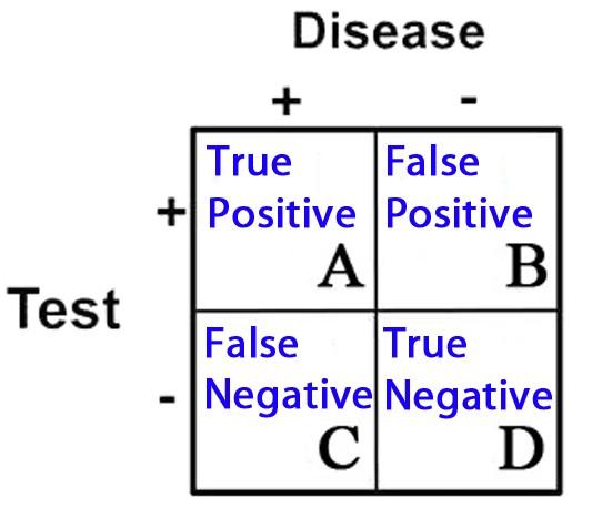
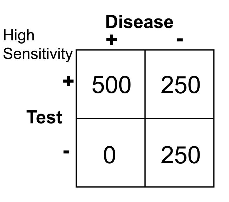
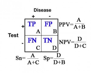
2 X 2 Contingency Table = A 2 X 2 (2 by 2) grid that is created to display the results of a comparison between two categories, e.g. an investigational subject (“test” group) and a gold standard (which determines, for example, who has and does not have disease). There are four possible results in a 2 X 2 table and they are as follows:

  1. True Positives = Subjects in the study that tested positive for a disease that in fact had the condition
  2. True Negatives = Subjects that tested negative for a disease that in fact did not have the condition
  3. False Positives = Subjects that tested positive for a disease that in fact did not have the condition
  4. False Negatives = Subjects that tested negative for a disease that in fact did have the condition

These categories are displayed in 2 X 2 grid as follows:

image35

Sensitivity (Sn) = In the group of patients that have disease (the “+” column under “Disease”), it is the proportion of patients with a positive test as it compares to all of the patients with disease. Specifically, it is the true positive rate as it compares to the sum of all of the true positives and false negatives.


Sn= (true positives)/(true pos’s + false neg’s)= A/(A+C)


In other words, sensitivity is the probability that the test result will be positive when the disease is present.

Specificity (Sp) = In the group of patients that do not have disease (the “” column under “Disease”), it is the proportion of patients with a negative test as it compares to all of the patients without disease. Specifically, it is the true negative rate as it compares to the sum of all of the true negatives and false positives.


Sp= (true negatives)/(true neg’s + false pos’s)= D/(D+B)


In other words, specificity is the probability that the test result will be negative when the disease is not present.

Positive Predictive Value (PPV) = The probability that the patient has disease when the test is positive (the “+” row for “Test”).


PPV=(true postives)/(true pos’s + false pos’s)= A/(A+B)


Negative Predictive Value (NPV) = The probability that the patient does not have disease when the test is negative (the “-” row for “Test”).


NPV= (true negatives)/(true neg’s + false neg’s)=D/(D+C)


Calculations for Sn, Sp, PPV, and NPV are depicted in the table below:

image36

Likelihood Ratio(LR) = This concept incorporates the use of sensitivity and specificity to determine the likelihood that a test will effectively yield the probability of the presence or absence of a disease state or condition. LR’s are utilized to provide a quantitative value in performing a particular test.

Number Needed to Treat (NNT) = The number of patients that need to receive a particular therapy until one is likely to experience benefit from this therapy: e.g.

the number of men over the age of 60 that need to take an aspirin per day until we see one less heart attack (compared to the population of men over 60 not taking aspirin).

Number Needed to Harm (NNH) = NNT is also used to measure harm. The number of patients that need to receive a particular therapy until you are likely to see one harmful event occur is called the Number Needed to Treat to Harm, or NNTH: e.g. the number of men over the age of 60 that need to take an aspirin per day until we see one patient sustain a bleeding stomach ulcer (compared to the population of men over 60 not taking aspirin).

Risk Ratio and Odds Ratio (RR and OR) = Both of these terms refer to the chance of an outcome between 2 groups. They are calculated similarly, but not exactly the same, and typically draw the same general conclusion. The Risk Ratio (RR), also called “Relative Risk,” is a percentage of chance. The OR is based on “odds” and not percent. These concepts will be described in greater detail later in this chapter and in the appendices.

Probability = In essence this is what statistics is all about: the likelihood or chance that a certain outcome will result. Probability is typically expressed as a fraction or percentage.

Population = Reference to all people, or at least all of those with a particular condition.

Sample = It is typically not possible or practical to study an entire population; for that reason, researchers typically study some proportion of individuals in this population. With appropriate sampling (i.e. elimination of bias), there is the greatest chance that the conclusion drawn will represent the entire population.

Bias = Whether intentional or not, it is the unfair favoritism of one particular outcome over another. Some of the common types of bias in medical literature are:

Selection Bias = typically the result of a non-randomized or poorly randomized study

Publication Bias = Pharmaceutical companies are frequently accused of this: e.g. only publishing “positive” studies, i.e. those that reveal their drug is beneficial. In contrast, they may not publish studies that are not favorable for their particular drug.

Surveillance Bias (aka. Detection Bias) = When one group of patients is

followed more closely in a study than another group. This typically happens because one group is considered to be more sick or more of interest by the investigators because they are the study group and not the placebo group.

Information (Recall) Bias = When information is gathered by way of the patients recollection of events, errors can occur because the patient may not recall pertinent details. This type of bias can also occur when there isn’t consistency in how questions are asked of patients: patients may give different answers depending on how they are asked questions. This is the classic, “garbage in, garbage out” error.

Spectrum Bias = Occurs when patients are studied at different points in their disease course: e.g. early vs. late appendicitis, patients have different likelihoods of positive findings on radiologic imaging.

Confounders = Results may be skewed in a study because of factors that the investigators didn’t account for. This type of error can give a false impression of cause and effect: e.g. immunization rates among kids have increased since the 1970’s and the rate of autism has increased since the 1970’s. Conclusion: Immunizations cause autism. In fact, there are many confounders: change in definition of autism to autism-spectrum disorder, better detection of autism, and many others.

Null hypothesis = The assumption that there is no difference between groups being studied.

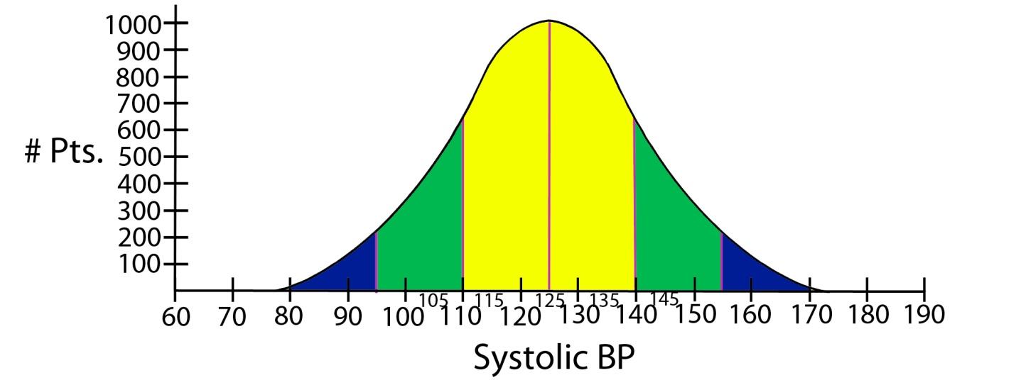
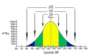
Normal distribution of data = The typical “bell shaped curve” that results when there is a normal distribution of data around the mean.

image38

Standard deviation = Quantifies the distribution of data around some average value: e.g. the second standard deviation from the mean identifies about 95% of values measured, and does not include the roughly 5% of measured values that remain (in medicine, these latter values would be considered to be significantly different from the mean or average).

image40

Statistical significance = The point at which, or the range of values outside of which, an event is not likely to have occurred by chance alone. Statistical significance establishes what is accepted to be a true difference between two groups (e.g. therapy 1 is better than therapy 2). Statistical significance is most commonly determined in one of two ways in the medical literature: either by p-values or confidence intervals.

p-value = p stands for “probability. It is the point at which statistical significance is defined. In other words, the point at which it is accepted that a difference between two groups is not due to chance. Traditionally the most common measure of statistical significance in medical literature, the p-value is typically set at 0.05. This means that there is a 5% probability (or less) that the results obtained in a study are due to chance alone: or stated another way, if the study were performed again, there is a 5% (or less) probability that the result would be outside the cutoff point of the p-value.

Confidence interval = In a comparison of populations, it is a range of statistical values (e.g. means, or LR’s) within which the “true result” is likely to reside. Confidence intervals are used to reveal the reliability of an estimate, and is typically set at 95%. This means that if the study were repeated 100 times, we would expect the result to fall within this range on 95 of those trials.

Bayesian Analysis or Bayes’ theorem = This is a form of deductive reasoning; that is to say, the clinician subjectively determines the probability of a particular event (prior probability), then performs a test that has some known likelihood of supporting or refuting that belief. In the end, then, the clinician draws a conclusion, posterior probability, and determines if the result is sufficient or if more testing needs to be done.

Explanation of Statistical Terms and Concepts Used in the Diagnosis of Disease:

Sensitivity and Specificity:

Arguably two of the most important statistical concepts that a physician (or other medical provider) needs to understand in order to perform a critical review of medical literature are sensitivity and specificity. In a perfect world with a perfect study, a test would be positive only when disease is present and negative only when there is absence of disease. Let’s imagine a study in which we are evaluating the reliability of a test to determine the presence of pancreatitis (inflammation of the pancreas) in 1000 patients with abdominal pain. Let’s imagine that the study results turned out as they are illustrated below.

image30

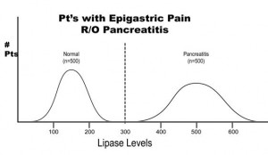
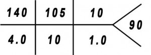
The results of this study reveal that there are 500 patients with abdominal pain that did not have pancreatitis and 500 patients with abdominal pain that did have pancreatitis (based on some kind of “gold standard” such as a CT scan). In this study, it was found that by using the investigational lab test, a lipase level, you could accurately diagnose pancreatitis when the level was greater than 300 units. Conversely, all patients with a level less than 300 were found to not have pancreatitis. This would be illustrated as below in a 2 X 2 contingency table.

image29

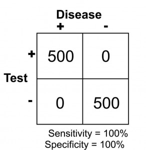
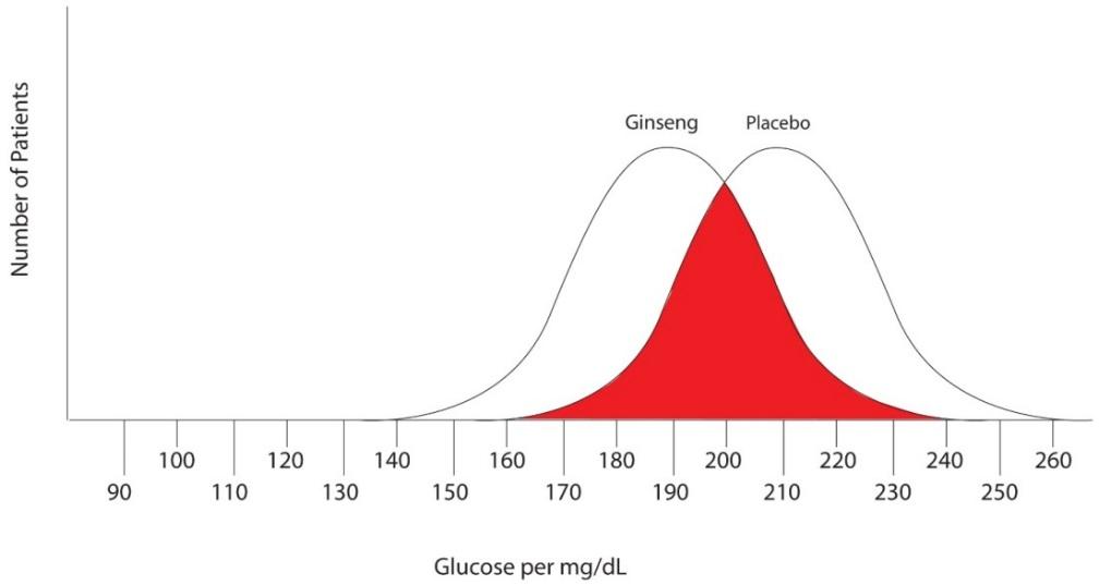
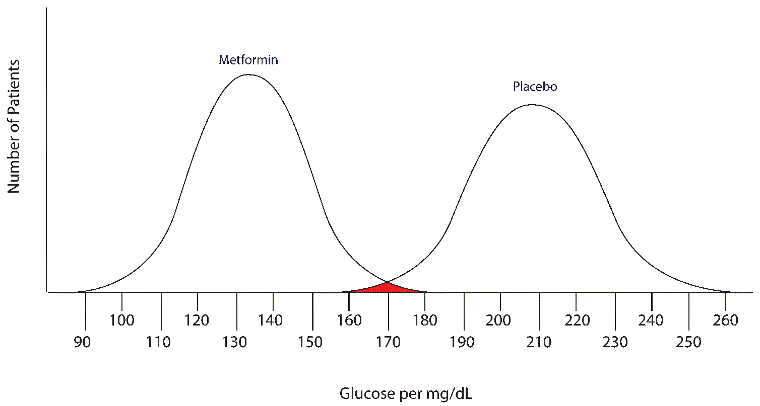
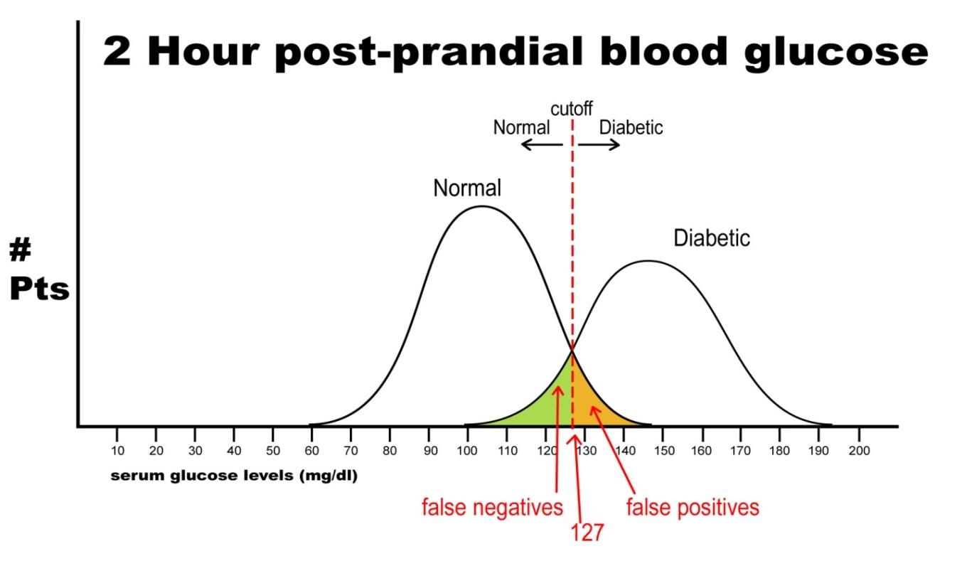
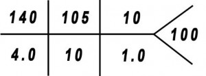
Note that there are no false negatives and no false positives, thus the test has 100% sensitivity and specificity. A more common scenario is the situation in which there is overlap of results, in that some patients without disease that have an extreme result will be found to have disease based on a test (false positives) and some patients with disease will be found to not have a positive test (false negatives). Imagine the study illustrated below in which investigators are looking to diagnose the presence of diabetes based on the results of a blood (serum) glucose level 2 hours after eating a standardized meal (postprandial glucose check). Imagine that researchers checked 500 people with and 500 people without diabetes (disease determined again by some accepted gold standard), and came up with the following results.

image32

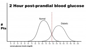
Notice that there is a wide range of glucose levels in both diabetic and non-diabetic patients at 2 hours after eating. Notice, as well, that there are cut-offs that define normal and abnormal. Where we choose these cut-offs to be determines the sensitivity and specificity of this test. To maximize both sensitivity and specificity, and get the overall best accuracy for this test, we choose a cut-off that is in the middle, where our two populations intersect.

image31

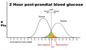
In this case, any patient with a serum glucose level greater than or equal to 127 (the middlemost value) at 2 hours postprandial would be labeled as diabetic, and any patient with a serum glucose level of less than 127 would be labeled non-diabetic. This would yield the following results:

image33

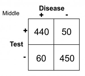
Sn = 440/(440 + 60) = 0.88 or 88%

Sp = 450/(450 + 50) = 0.90 or 90%

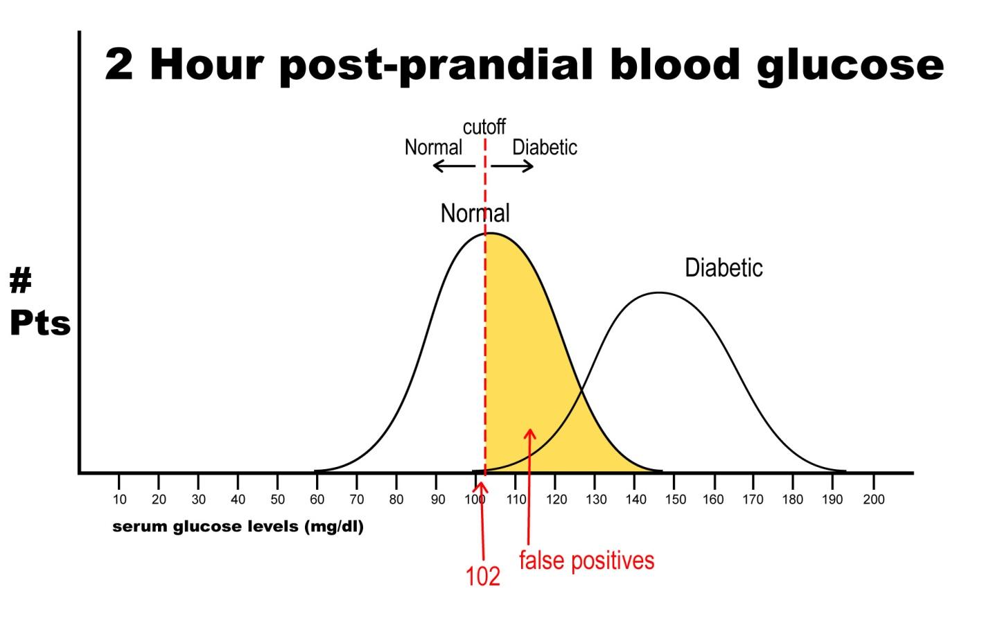
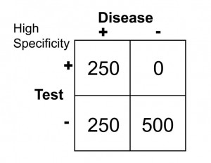
Let’s say, now, that it is vital that this test be 100% sensitive so that we do not miss any patients that could potentially have diabetes. If we move the cut-off to a glucose level of 102, then we get the following results:

image41

Notice that there are no false negatives and thus 100% sensitivity. Notice as well, however, that the number of false positives has increased dramatically. This change in cut-off value would yield the following results:

image42

Sn = 500/(500 + 0 ) = 1.0 or 100

Sp = 250/(250 + 250) = 0.50 or 50%

Notice that by choosing this cut off point the test does exceedingly well at identifying patients with disease. In changing to this cut off point, however, the number of false positives increases substantially, so we are now going to identify many people as having disease when they really do not have it (has low specificity). Notice further, though, that because a highly sensitive test has a low false negative rate, when the test is negative, it virtually rules out disease (i.e. a negative result in a highly sensitive test means the patient does not have disease).

Finally, let’s imagine that we need to have 100% specificity with this screening test because we are going to expose the patient to a treatment that could be deleterious to non-diabetics. If we change the postprandial glucose cut-off to a level of 148 as the definition of diabetes, then we get the following results:

image43

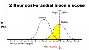
In this case, there are no false positives, but 250 false negatives. This yields the following results:

image44

Sn = 250/(250 + 250) = 0.50 or 50%

Sp = 500/(500 + 0) = 1.0 or 100%

This selected cut off point makes this test exceedingly good at identifying patients without diabetes, but it is also labeling a lot of people as non-diabetic when they really have disease (the false negative rate increased as we increased the specificity). Notice in this case, though, that when the test is positive in a highly specific case that this virtually assures that the person does have disease: i.e. when the person’s blood sugar, in this example, is greater than 148 mg/dl, the person is essentially certain to have diabetes.

Positive and negative predictive values:

As stated previously, PPV is the probability that the disease is present when the test is positive, and the NPV is the probability that the disease is not present when the test is negative. With PPV and NPV, we are now dealing with how well a test performs. Predictive values, however, can change dramatically as the prevalence of disease changes from one population studied to another. If bias and confounders are not introduced, sensitivity and specificity do not change from one study to another despite a change in prevalence of disease. To illustrate this point, imagine the following study results.

A health system studies the efficacy of having housekeeping staff collect throat swabs on patients, as a cost saving measure, instead of having nurses perform the swabbing of the patient’s throat (the gold standard). Investigators perform 2 studies evaluate patients for Strep throat. In the first study, investigators enroll patients that have a sore throat, fever, and exudate (pus) on the tonsils. In this study, 50% of patients are found to have Strep throat by the “gold standard.” In the next study, investigators evaluate patients with sore throat, fever, and no exudate. In this case, using a gold standard, the prevalence of disease (proportion of patients found to have Strep throat) is found to be only 1%. The sensitivity this test when housekeepers obtain the specimens is 20% and the specificity is 95% (meaning that the housekeeping staff are not too good compared to nurses when it comes to swabbing the tonsils for Strep, but when they did swab the tonsils they did it right in that they did not have many false positives). In both cases, the Sn and Sp are the same, 20% and 95% respectively. The results obtained are below. Before looking at the answer, calculate the PPV and NPV for each study.

image47

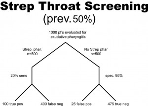
PPV = 100/(100 + 25) = 0.80 or 80%

NPV = 475/(475 + 400) = 0.54 or 54%

image48

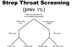
PPV = 2/(2 + 49 )= 0.039 or 3.9%

NPV = 941/(941 + 8) = 0.99 or 99%

So, what can we glean from these studies? Well, first of all, the housekeepers seem to have a very poor ability to obtain proper specimens compared to nurses (if we accept their results as the gold standard) since the sensitivity is only 20% in each study (i.e. there are a lot of false negatives). In other words, this study reveals that patients with disease were not diagnosed 80% of the time because the specimen was not collected as well as if it had been collected by the nurse, but why would we expect a housekeeper to know how to do this task? Notice, though, that the PPV for the test is actually pretty high, at 80%, when there is a high prevalence of disease. That is because PPV tends to track with Sp as they both share “false positives” in their calculation. Notice that the NPV is actually pretty poor when there is a high prevalence of disease, but it is very high when there is a low prevalence of disease. Can you see why this is the case? Notice that with a prevalence of only 1%, 99% of patients do not have disease. That being the case, if you didn’t do the test at all and just picked a patient randomly from this population as not having disease, you’d be correct 99% of the time. We can conclude for this study that having the housekeepers obtain specimens for Strep screening appears to be a bad idea.

Likelihood Ratio (LR)

Likelihood ratios are used when assessing the likelihood of a disease being present based on a certain test. Sensitivity and specificity are used in the calculation of LR’s. By helping to determine the usefulness of a test, LR’s predict the chance that a particular disease state exists. LR is a term commonly seen in modern medical literature because it provides very practical and useful information as you look to counsel individual patients regarding a particular test.

LR+ is for “ruling in disease” (determining disease is present).

LR- is for “ruling out disease” (determining disease is not present).

The calculations are as follows:

LR+ = sensitivity/(1-specificity)

We call this LR+, or positive likelihood ratio, because it is the likelihood that the person has a particular diagnosed condition. There is also a negative likelihood ratio, or LR-, which indicates the likelihood that someone does not have a particular condition. It is calculated by means of the following:

LR- = (1-sensitivity)/specificity

Likelihood ratios yield results that range from 0 to infinity:

LR range: 0———-1———-2———-//———-10—-infinity

Whether talking about LR+ or LR-, if the LR value is 1, it means this is a neutral value. That means it gives no indication as to the usefulness of the test. If a study is done where LR+ = 1, that means that when the test is positive the patient is just as likely to have disease as not have disease. If the calculated LR+ is found to be greater than 5 but still less than 10, the test in question is considered to be moderately useful in its predictive ability for determining the presence of disease. If the LR+ is greater than 10, it is considered very useful in determining the presence of disease. If the LR- is calculated to be less than 0.2 (1/5), then the test is considered moderately useful in determining the absence of disease. If the LR- is less than 0.1, it is considered very useful. The higher the LR+ is and the lower the LR- is, the more useful the test is. So, to interpret the value of LR+, for example, if a test is found to have LR = 5, that means that a patient with a positive result is 5 times more likely to have the disease being tested than not to have the disease. For more explanation about LR’s, and for more examples and calculations, see appendix 5a.

Case scenario

A 50 year old woman presents to your internal medicine office with a headache. She has had this waxing and waning headache for the past 12 weeks and is concerned because she has a friend that had a similar headache and the “Lyme disease specialist” that she saw determined it was the result of chronic Lyme disease. The patient wants to receive antibiotics to treat this presumed Lyme related headache. The patient doesn’t want to waste her time with testing for Lyme because, “the tests are inaccurate.” In your evaluation of her you discover that she does spend a fair amount of time in the outdoors but hasn’t been camping and doesn’t recall ever having a tick bite or a rash. You inform her that you think she is at very low risk for Lyme disease and thus unlikely to have any kind of Lyme related headache. You concede that the accuracy of the test is not perfect but that the CDC (Centers for Disease Control) reports the test to be 80% sensitive and 90% specific for diagnosing Lyme disease. Should this patient receive antibiotics for Lyme disease without any testing? If a Lyme test is performed and found to be negative, what is the negative predictive value of this test?

Other Clinical Integumentary Scenarios

The following scenarios are designed to walk the future physician through a variety of real life situations that highlight disorders of the integumentary system. Read the introduction closely and work through the scenario to ultimately diagnose and treat this patient’s condition.

Clinical application: Our burn patient

In the emergency department, oxygen is being given by face mask and an intravenous line has been inserted. Our patient has pale and charred skin on the scalp, face, torso, arms and hands, and upper legs. She is spared any burns below the knees and there are small areas of sparing on the torso. After a brief examination, you order some potent intravenous pain medication. At the same time, you prepare for ambulance transportation to a specialized burn unit, which in this case is eighty miles away.

There are many issues that need to be addressed and treated before transport occurs. She first needs her airway and breathing protected and she needs pain control. This necessitates endotracheal intubation and assisted ventilation. Large doses of narcotic pain medications are given. Recall, as well, that one of the major consequences of major burns is fluid loss. In our Renal chapter we learned that we use an isotonic solution such as “normal saline” to expand the intravascular space and that is what we will use for fluid replacement here. Our colleagues at in have developed a formula for fluid resuscitation in burn victims and it is as follows: take the percent body surface area that has sustained second and third degree burns, multiply this number by the patient’s weight in kilograms, and then multiply this product by 4 ml. This is how much fluid that the patient needs to receive in the first 24 hours. Her burn is 80% BSA and her weight is 50 kg, therefore she needs to receive 16 liters of isotonic fluid in addition to her maintenance fluid in the first 24 hours.

Parkland Formula for fluid resuscitation in burns:

24 hour fluid requirement = weight (kg) X %BSA of burn X 4 ml/kg

24 hour fluid requirement = 50 kg (80) (4ml/kg)

24 hour fluid requirement = 18.1L

Understandably, the girl’s family is stunned by the event, and wants to know why she can’t stay at their local hospital. They also want to know if she will be okay and if she will have any long term problems. You provide an accurate yet compassionate assessment of the situation.

Reviewing the pathophysiology and treatment of burns above, it is clear that this patient has a major life-threatening burn. In a young healthy patient, survival is possible but certainly not guaranteed. You discuss with the family the need for highly specialized care of the burn itself, including dressing and skin grafting, along with the intensive care to deal with the fluid losses and severe infections that commonly accompany these injuries. In addition, you discuss the need to place an endotracheal tube prior to transfer in order to protect the airway and permit more aggressive sedation and pain control. And yes, there will be scarring, but skin grafts and the use of cultured skin, as well as reconstructive procedures will go a long way towards restoring an acceptable cosmetic outcome. This event will change her life in many ways, but she will still be the same wonderful person she was before.

Pathophysiology & Treatment of Burns

A burn is an injury to the skin that occurs as a result of excessive heat (thermal burn) or as a result of exposure to a chemical (typically a strong acid or base). Burns are categorized by the “degree” in which the skin is damaged, and by the percentage of body surface area involved. We will focus on thermal burns as it relates to our patient scenario. In addition, the treatment of thermal burns is straightforward. Thermal burns are generally broken into 3 categories: first degree, second degree, and third degree.

A first degree burn is a superficial burn, involving only the most superficial layer of skin: the epidermis. Mild sunburn (no blisters) is a good example (Figure 6). The skin is red, blisters are absent, and important structures are spared. While these injuries can be painful, healing without long term consequences is the expected result, even in a burn involving virtually 100% of the body surface area.

image17

Figure 6 Cutaway view of the skin: first degree burn. The injury is limited to the superficial layers of epidermis and dermis.

Second degree burns (Figure 7) involve deeper layers of the skin. Superficial layers separate and form fluid-filled blisters. In a large burn, more than 15 – 20 % of body surface area, enough body fluid can be diverted into these blisters to cause hypovolemic shock. When the blisters rupture, the area becomes susceptible to infection. Because the deeper layers containing the nerve endings are spared (not damaged), second degree burns are very painful. With partial sparing of the growth layer, most of these burns will heal with new skin rather than scarring.

image18

Figure 7 Cutaway view of the skin: second degree burn. This injury involves deeper layers of the dermis. The deepest layers and important structures – nerves, blood vessels, sweat and sebaceous glands and hair follicles — remain intact. Remnants of the germinal layer remain intact, conferring potential for healing by growth of new skin.

Third degree burns (Figure 8) destroy the entire thickness of the skin. Because nerve endings are destroyed, the burn is painless, but the consequences are more severe. The germinal layer of the epidermis is destroyed in this full thickness burn; therefore the body is incapable of growing new skin in the affected area.

image19

Figure 8 Cutaway view of the skin: third degree burn. This is a full-thickness injury. Nerves are destroyed, so the wound is painless. The growth layers of the dermis are also lost, so healing will occur by scarring rather than by growth of new skin.

Fluid loss and infection can be life-threatening and victims of a major burn may spend weeks or months in the hospital. Among patients sustaining a severe burn to 80- 90% of the total body surface, only half will survive.

Because the burn heals by scarring rather than re-growth of skin, those who do survive are deprived of the protective, heat-regulating, elastic tissue that constitutes an important interface between us and our environment.

That brings us to the next issue regarding burns and that is the amount of skin involved. Besides depth, the other crucial factor regarding burns is how much surface area of skin has at least second or third degree burn involvement: this is referred to as the percent of total body surface area (BSA). Estimating this can be difficult but a good method is to use the “rule of 9’s” (Figure 9). For smaller burns we use the patient’s palm (not including the fingers or wrist) to estimate how many “palm sizes” a particular burn is. Each palm size is nearly 1% of BSA.

image14

Figure 9 The “Rule of 9’s”. The numbers are added to determine the percent of total body surface area that is burned.

Treatment of Burns:

Simple burn care consists of cleaning and dressing the wound. The biggest concern for the patient, however, is pain control. There is nothing like the pain of a severe second degree burn, and there is nothing like the instant relief one achieves by simply running the affected area under cold water or placing the burned area in a cool water bath.

image15

Figure 10 This is a deep second degree burn, the result of a lighter that exploded in this poor gentleman’s hand. Opiates are the mainstay of medications for pain control, after cool water. Anti-inflammatories (e.g. ibuprofen or naproxen) are also used as an adjunct for pain control. After reasonable pain control is achieved, dressing the wound with antibiotic ointment applied under a gauze dressing would be appropriate. The patient needs a one day follow-up to confirm the depth of the wound: which often looks more superficial on the first day.

When dealing with serious burns involving large surface areas of skin, it should come as no surprise that airway and breathing issues are high priority, as they are in all medical emergencies. Swelling in the throat, face, and neck can compromise the airway. Smoke inhalation can severely compromise the ability of the lungs to oxygenate blood, and loss of elasticity of skin over a chest burn can prevent normal chest expansion with inspiration. For these reasons, supplemental oxygen is commonly necessary, as is endotracheal intubation and assisted ventilation.

Hypovolemia due to fluid loss through the burn is the next major priority. Patients with large areas of third degree burns may require many liters of intravenous fluid every day for the first few days in order to replace these fluid losses.

While the region of the third degree burn itself isn’t painful, it typically coexists with areas of first and second-degree injury: both of which are very painful. Burn patients require large doses of strong pain medication.

Care of the burn itself is, surprisingly, not an early priority in major burns, but is crucially important once the ABC’s have been addressed. While less serious burn wounds can be treated with antibacterial ointments and bandages, larger ones require cleaning in whirl pools, coverage with more specialized dressings, and grafts from the patient’s own skin and/or cultured skin (grown in the laboratory).

Clinical Application: Our acne patient

Recall that our patient, a 15 year old girl, has moderately severe acne and is suffering emotionally because of this. Obviously we need to help this girl by treating her acne, but let’s take a moment to address her psychosocial issues (the girl’s ability to cope with her disease). Why did this girl go into such a serious downward emotional and social spiral from such a benign disorder? After all, acne is just pimples and it should be a simple problem to assess and treat – shouldn’t it? The answer is no, acne is not always a simple problem and it can leave both physical and emotional scars. Acne commonly occurs on highly visible areas such as the face, upper chest, and upper back, and can greatly affect self-esteem as it tends to disfigure these areas.

Regarding the patient’s acne, she has already tried a variety of soaps and even cleansers containing benzoyl peroxide. She is not seeing much success with this “first line” treatment. In an effort to minimize potential adverse effects, yet give her the best chance for success, what treatment plan would you recommend at this point?

Recall that estrogens (a major component of OCPs’) tend to suppress sebum production and frequently lead to an improvement in acne. Oral contraceptives could potentially regulate her menstrual cycle as well as prevent an unwanted pregnancy. Discussing the risks and benefits of this therapy would be prudent.

In addition to OCP’s, you will want her to continue using the antibacterial soap and benzoyl peroxide that she is already using. Another therapy to consider is antibiotics. You could start with topical antibiotics but most clinicians would probably go right to oral antibiotics with this particular patient.

“Surgical” treatment such as comedone extraction could be considered at this time but injections with anti-inflammatories and laser and dermabrasion would not be indicated this early in the treatment of this disease. Treatments like these are expensive and medical insurance plans offer minimal coverage for procedures considered “cosmetic” — performed primarily for reasons of appearance.

You discuss the treatment plan with the patient and her mother. You assure the girl that you are taking her acne problem very seriously and that you are going to see her back in just a few weeks to be certain she is doing okay. She knows that even if her acne is not controlled at that point that you have other therapies to offer. Both mother and patient leave feeling pleased.

Just then, a call comes in from the emergency department. They have just admitted a 12 year old girl with burns estimated to cover 80% of her body. She had been at a friend’s for a sleepover. They built a fire in order to roast marshmallows. New wood thrown on the ash wouldn’t light again this morning, so the patient grabbed a small can of gasoline and poured it on the fire. Immediately the flames rushed up the stream of gas, exploded gas into the air and on the patient, and ignited her hair and clothes. She ran to the house, and the father of the girl’s friend, hearing the commotion, ran to the patio to see what the matter was. He grabbed the young girl, now engulfed in flames, and rolled her in the grass to extinguish the blaze.

In the emergency department you find the girl to be quite stoic but clearly in pain. She is breathing on her own and is not coughing. She has pale and charred skin on the scalp, face, torso, arms and hands, and upper legs. She is spared any burns below the knees and there are small areas of sparing on the torso. What do you do?

Pathophysiology & Treatment of Acne

There are actually a number of types and classifications of acne and related conditions, but for this discussion we will concern ourselves with acne vulgaris (vulgaris = common). As the name implies, this condition is encountered frequently but it is anything but simple. Among the causes of acne are hormonal influences, genetic predisposition, contact with skin (e.g. touching face with dirty hands or use of chin straps on helmets), and use of certain medications.

Interestingly, particular foods like potato chips and chocolate, long thought to be a cause of acne, have not widely been associated with inducing this skin disorder. Among its consequences are the development of more serious skin infections, scarring, embarrassment, social isolation, and other psychological trauma. Acne treatment has become a multi-billion dollar industry.

Typically, acne starts when the sebaceous glands are induced to produce more oil. This commonly happens during puberty, when levels of sex hormones, especially testosterone, rise. (Testosterone is usually considered a male hormone, but females have small amounts of this as well). Under the influence of these hormones, sebaceous glands enlarge, and the increased sebum mixes with sloughed lining of the follicle and gets clogged at the neck of the hair follicle. This plug is the typical early mark of acne, the comedone. When the pore remains closed with the sebaceous plug, this is referred to as a closed comedone, commonly called a “white head.” When the orifice of the plugged pore enlarges as cellular debris accumulates and darkens, an open comedone or “blackhead” develops. As bacteria propagate behind this obstruction and in the rich environment of sebum, inflammation occurs and a papule develops. Further inflammation and the production of pus, from the body’s immune response to the infection, leads to the formation of pustules (Figures 3 and 4a-e).

image07

Figure 3 Progression of an acne lesion: an overview (Used with permission of Galderma Laboratories, Fort Worth, TX)

image08

Figure 4a Normally functioning hair follicle and sebaceous gland (Used with permission of Galderma Laboratories, Fort Worth, TX)

image09

Figure 4b Development of the microcomedo: the neck of the hair follicle becomes plugged by skin debris (from excess keratinization – sloughing of the outer layer of the lining of the follicle) mixed with sebum (which is produced in excess). (Used with permission of Galderma Laboratories, Fort Worth, TX)

image01

Figure 4c Progression of a closed comedone (“whitehead”) to an open comedone (“blackhead”). This occurs as the orifice of the pore enlarges and accumulates additional debris. (Used with permission of Galderma Laboratories, Fort Worth, TX)

image00

Figure 4d Progression from comedone to papule, resulting from infection and inflammation (Used with permission of Galderma Laboratories, Fort Worth, TX)

image03

Figure 4e Further progression from papule to pustule, with pus visible above the skin surface. (Used with permission of Galderma Laboratories, Fort Worth, TX)

image02

Figure 4f Papular and early pustular acne

When a hair follicle gets plugged, sebum accumulates behind the obstruction because the sebaceous gland keeps producing this oily substance. One particular bacterial species, Propionibacterium acnes (P. acnes), flourishes in this environment. The human body recognizes the rapid proliferation of bacteria in this sebum filled, occluded follicle and sends white blood cells (WBC’s) to fight this infection. These WBC’s release a variety of inflammatory mediators and this results in redness and swelling. As more WBC’s are drawn into the follicle they, along with the bacteria they kill and other cellular debris, accumulate as pus. As more pus develops within the follicle, the surrounding area becomes more reddened and swollen and even warm and tender. The resulting pustule is commonly referred to as a pimple. The longer and more intense this inflammatory reaction is, the more the lesions progress. Though small at first, these pustules can grow to become large cysts, and these cysts can coalesce and form fistulous tracts between them and result in severe deformity and scarring of the involved areas of skin (Figure 5a-g).

image04

Figure 5a Hair follicle and sebaceous gland: early accumulation of sebum (yellow). The follicle is intact. The small bubble insert shows an enhanced microscopic view of the follicle lining.

image10

Figure 5b As pus (white blood cells and bacteria) accumulates within the follicle, pressure rises and the wall of the follicle begins to bulge.

image11

Figure 5c When the pressure within the follicle reaches a critical point, rupture of the follicular wall occurs and sebum, white blood cells, and bacteria are released into the subcutaneous tissue. This attracts scavenger cells known as macrophages (green) to help clean up this infection.

image12

Figure 5d Common acne cyst

Unfortunately, acne commonly affects some of the most visible areas on the body. Scarring is the worst (physical) long term consequence of acne (Fig. 5f). In the past it was often unavoidable, but now there are a variety of treatments that may control the acne before it reaches the scarring stages, as well as procedures that can restore scarred and irregular skin to a more normal appearance.

image13

Fig 5e Another acne cyst in background of more serious cystic acne

image16

Figure 5f Scarring as a result of severe cystic acne

Treatment of Acne:

As in all science-based medicine, therapeutic interventions are based, whenever possible, on an understanding of pathophysiologic processes. Layered on top of this is knowledge of the specific treatments, both their expected results and potential adverse effects, as determined by clinical research. Finally there is application to the individual patient ideally based on collaboration between patient and practitioner.

In the early stages of acne, simple treatments are often all that is needed. Antibacterial soap is one of the simplest first treatments: it kills the offending P. acnes bacteria and it cleans off excess oil (sebum) from the surface of the skin. Benzoyl peroxide, which can be purchased without a prescription, is another first line treatment. This agent is available in a variety of liquids and gels and mixed in a variety of media allowing it to dry the skin, dissolve comedones, and break down excess sebum on the surface of the skin so it can be easily washed away. It also has some antibacterial activity and is combined with other agents in an effort to make it even more effective. This sort of treatment carries little risk of unwanted effects so it is perfect for an initial treatment. In minor acne this treatment may be all that is necessary.

If these agents are not sufficiently effective, other types of topical treatment can be considered. Derivatives of vitamin A (retinoids) cause shedding of skin cells and reduce the accumulation of excess keratin within the follicle. Topical antibiotics are another second line of defense against acne and they work by decreasing bacterial proliferation. These two agents are commonly used in combination in moderate acne. Both of these agents however, carry some risk of significant skin irritation and allergic reaction.

If the acne reaches the nodular or cystic stage, more aggressive treatment may be considered. Direct care of the lesion by the dermatologist – extracting the sebaceous plugs from comedones or injecting nodules and cysts with anti-inflammatory medications can be helpful in the short term, but they are time-consuming and expensive.

In addition to the topical agents described above, oral medications may be needed as well. Both antibiotics and retinoids come in oral forms, and tend to be more effective when taken in this way. Introducing a drug into the bloodstream, however, generally introduces the potential for more serious side effects as well. The retinoids especially carry a number of known risks, including liver toxicity, sores at the side of the mouth and elevation of undesirable lipids in the bloodstream. The most significant issue with these drugs is that, when taken during pregnancy, they pose serious dangers to a developing fetus by causing birth defects. Since young women in their childbearing years are among the most common types of patients seeking treatment for a cosmetically significant condition, this issue has resulted in strict formal protocols to ensure that these medications are not administered during pregnancy.

And speaking of pregnancy, recall the role of the sex hormones in the production of sebum: testosterone tends to increase the quantity and density of sebum, while estrogens have the opposite effect. Hormonal manipulation, usually by the administration of estrogen as an oral contraceptive, can have a beneficial effect for the patient with acne. For younger women who may desire the contraceptive and menstrual period-regulating effect of birth control pills, this may be a good solution. For women ready to have children, this would not be a good option. Moreover, even such “natural” substances as estrogens can be associated with increased risk of blood clots in the legs, lungs and brain, and possibly some cancers. Hormone therapy is obviously not a viable choice for males since an excessive influence of estrogen would result in gynecomastia (development of breasts) and other undesirable effects.

Finally, what can be done for the patient who, after years of severe acne, ends up with the scarring and uneven skin surface? Fortunately some of these changes can be erased, or at least improved, by processes designed to very carefully eradicate the superficial layers of damaged skin. This is carried out by the use of laser light, chemicals, and abrasives. The expectation is that the healing process will result in a smoother and more uniform appearance to the skin.

Background, Anatomy, Physiology

Skin has many functions that are necessary for survival: it serves as a barrier to infection, it stores and holds fluids and energy, it plays a large role in regulating body temperature, and it contains sensors that allow us to feel the environment around us.

As the largest and most visible organ of the body, skin also plays a prominent role in the perception of physical beauty. Diseases of the skin can lead to embarrassment, loss of self-esteem, and social isolation. Some will deal with debilitating skin disorders better than others. As a healthcare provider, you need to individualize treatments for skin disorders based not only on the patient’s physical condition, but also on the patient’s emotional response to the severity of the condition. Treating acne, for example, seems straightforward but a provider can easily miss the emotional importance of this condition. Conversely, it is not difficult to be lured into aggressively over-treating this disease and lose sight of the fact that there are risks to some therapies that may outweigh the benefits.

Anatomy and Physiology of the skin:

In the average adult, the skin covers an area of almost 2 square meters. Cutis is the Latin word for skin and thus when we refer to structures within the skin we refer to them as “cutaneous” (of the skin) structures. Skin is composed of 2 major layers: the epidermis and the dermis (Figure 1).

image05

Figure 1 Cross-section of the skin: epidermis, dermis, and subcutaneous tissue

The epidermis is the outermost layer of the skin. This is the body’s first defense against external threats such as desiccation, extremes of temperature, ultraviolet radiation, and microorganisms. The epidermis is thin and primarily consists of multiple layers of dead cells that had their origins in the deeper, generative layer. At the base of the epidermis is the basal layer. This single layer of cells is made up of keratinocytes (squamous epithelial cells that form the outer skin and produce keratin) and melanocytes (pigment producing cells that determine skin color). It is the only place in the epidermis that proliferation (cell division for new cell growth) takes place. Beneath the epidermis, the dermis is a more complex layer housing a variety of structures important to the functions of the skin as outlined above (Figure 2). Sweat glands open and close in response to stimuli from nerves and hormones; they help to maintain a constant body temperature. Hair follicles are genetically programmed to produce various types and quantities of hair in different parts of the body and are supplied with the tiny muscles that “make your hair stand on end.” This phenomenon, in other animals, serves to increase the insulating ability of the hair (fur), but in humans is mainly responsible for the phenomenon of “goose pimples.” Closely associated with the hair follicles are the sebaceous glands which secrete the oil that adds to the waterproof properties of our skin. Rather than being evenly distributed over the skin, the sebaceous glands are concentrated in the head, neck and upper torso, which has medical consequences that will be described below.

image06

Figure 2 A small block skin and a depiction of the structures within: nerves, blood vessels, sweat glands, sebaceous glands, hair follicles

Below, or deep to, the dermis is the subcutaneous tissue, composed largely of loose connective tissue and fat. This layer may be considered part of the same functional unit as the dermis and it is physically attached to the skin more than to the underlying tissues. The dermis and subcutaneous tissue serve as the primary insulation of the body.

Having their origins elsewhere, but traveling through the subcutaneous tissue to their destination near the surface of the skin are numerous blood vessels and nerves. The second-smallest branches of the blood vessels, the arterioles, can constrict or dilate to regulate flow of blood into the skin’s capillary bed in order to expose more or less surface to the environment and serve to regulate body temperature. The nerves terminate in specialized endings that serve as transducers for the senses of touch, vibration, and hot and cold.

Pathophysiology of the Integumentary System:

There are a variety of diseases and injuries that can affect our skin. In the paragraphs below, we will highlight a broad range of these conditions, but with a special emphasis on acne and burns.

Because the skin is a major interface between ourselves and our environment, it should not be surprising that the environment can have an impact on the skin. We refer, of course, to trauma of various sorts: lacerations (cuts), abrasions (scrapes), contusions (bruises), and burns would describe most types of injury to the skin. Loss of major areas of skin – for example, a deep burn of more than 40% of the body’s surface – can be life-threatening. Fortunately, in all but the most serious injuries, the skin has a remarkable ability to regenerate and heal, often with little need of medical intervention.

Infections of the skin reflect the interaction of skin with the microbial world outside. While bacteria, viruses, and fungi often live within the epidermis, the term infection connotes an invasive process in which large populations of microbes breach the skin’s defenses. When the skin is invaded by pathogens such as these and the invading organism begins to multiply, it is referred to as cellulitis. This infection can result in destructive processes that progress to loss of a body part or even death. Host defense mechanisms, such as inflammation and fever, also come into play. White blood cells invade the area and form the pus in abscesses and tiny pustules in some skin infections.

Inflammation is a complex phenomenon in which the body’s defense mechanisms produce characteristic changes such as localized warmth, redness, and swelling. While inflammation may be an appropriate reaction to infection and other injuries, it may also occur when it is not wanted. The generic term for inflammation of the skin is dermatitis and there are dozens of kinds skin diseases such as eczema, often associated with allergies and asthma. Poison ivy is an example of a contact dermatitis. In this case, a resin from the plant penetrates the outer layer of skin and the response by the body’s immune system leads to itching, inflammation, and blisters. There is a wide range of skin diseases in which inflammation is a prominent feature. In some cases the inflammation can be so severe as to result in major skin loss and even death.

Several skin problems are the result of a dysfunction of hair follicles. Acne (discussed in detail below) is the most common of these disorders, but there are many other conditions in which the hair follicle gets closed and allows for the development of inflammation and infection within the follicle. These conditions include pilonidal abscess (infected hair follicle that develops into an abscess and is located in the upper central crease between the buttocks), hidradenitis suppurativa (inflammation and abscesses that develop in the folds under the arms and in the groin), and hot tub folliculitis (inflammation that occurs in hair follicles after being in a hot tub and reacting to chemicals and/or infection).

Like any other organ in the body, skin is susceptible to abnormal growths and cancer. Non-cancerous growths are referred to as “benign”, meaning they are biologically programmed not to spread beyond a limited area. Cancerous (malignant) tumors invade local tissues and can spread to other parts of the body. Cancer of the skin is the process in which a germinal cell begins to divide in an unusual manner and at an accelerated rate. This leads to a bump or colored area on the skin. Some cancers grow as a localized lesion (e.g. basal cell carcinoma) until they become very large and others begin to spread (metastasize) throughout the body. One particularly serious form of skin cancer is malignant melanoma, recognized by its dark coloration. Skin cancers, and even benign growths, are often the result of excessive UV exposure from the sun.

In contrast to the cancers, some cutaneous diseases are the result of abnormal growth of normal tissues. Psoriasis is a disorder in which the skin simply grows too fast in some areas and becomes thickened with too much of the outer keratin layer (hyperkeratinization). This is a benign growth but can be very debilitating and disfiguring.

Because the skin is the most visible component of our bodies, many systemic diseases can be diagnosed by their unique prominent dermatologic manifestations. It is common knowledge that varicella (chickenpox) causes a rash consisting of tiny blister-like lesions (vesicles) on a red base. Measles and rubella, now uncommon in the U.S. due to vaccination, cause rashes with classic characteristics: a doctor only needed to see the rash, years ago when it was common, to diagnose this disease. There is a wide assortment of other systemic infections, allergic conditions, and inherited diseases that are accompanied by characteristic skin changes.

Dermatological Definitions:

In all medical specialties, a localized abnormal structure in the body is termed a lesion. Because the field of dermatology is heavily weighted towards the visual, a fairly precise system of terminology has been developed to describe lesions of the skin. For example, a small, non-elevated colored spot is called a macule; when the spot is elevated, it becomes a papule. A tiny superficial fluid-filled lesion is a vesicle, while a much larger one is a bulla. A papule is a small, raised, conical shaped lesion, commonly red and inflamed. When a papule becomes slightly larger and fills with pus, it is called a pustule. A soft, red, raised, blanching (turns skin color when compressed) skin lesion that is pruritic (itchy) and can be quite large with irregular borders is urticaria (hives): typically the result of an allergic reaction. These are not entirely arbitrary categories, as location within the skin structure and specific pathophysiology confer on these lesions their characteristic appearances.

Patient Scenario

As a family physician in a small community, your day started like any other as you went to see the first patient in your office at 8:05 AM. Sitting across from you is a 15-year-old girl for whom you have been caring since birth, but haven’t seen for more than 3 years. The young girl is bent over with her face in her hands, and her mother is tearful with an arm draped around her daughter. The mother states that she just found out last night that her daughter has not been going to classes regularly for almost two months, and has been classified as truant. Previously an honor student, the patient has begun to fail in school and has been ostracized from her group of high-achieving friends. In the interview, you learn that the girl has been developing increasing anxiety and depression over the past couple of years. She has little self-esteem and refers to herself as “ugly.” She raises her head up as she says this and reveals what she says is the source of “all” of her problems. She states that she has been made fun of at school and that nobody likes her. When you closely examine the girl’s face and back, you see numerous pustular and cystic acne lesions, and even several small, pitted scars. This is a moderately severe case of acne. What do you do? Jot down a few ideas of what you think the issues are and what you would do to help this troubled girl.

Acid/Base Pathophysiology

There are 4 basic acid/base disorders: 2 metabolic disorders (metabolic acidosis and alkalosis) and 2 respiratory disorders (respiratory acidosis and alkalosis). These disorders can, for the most part, be explained by the using the carbonic acid equation:

image01

Disorders that move the carbonic acid equation to the left, or result in greater H+ concentrations, cause acidosis. The converse is true, in that disorders that move the equation to the right, or result in a lesser H+ concentration, cause alkalosis. There are two exceptions to this rule: 1) Drinking lots of water (adding substrate to the right side of the equation) does not result in any significant acidosis, and 2) A disorder resulting in increased H+ will move the equation to the right and HCO3will be used up, but there is still an increase in H+ so there is acidosis, not alkalosis. Each disorder will be discussed individually.

Respiratory acidosis:

Respiratory acidosis results from an excess of CO2 in the serum. This typically occurs because normal air exchange is impaired at the level of the alveoli. Causes may be that the patient’s ventilations are suppressed, as can be the case with an opiate overdose, or that there is airway obstruction impeding adequate flow of air in and out of the alveoli, as can be the case with asthma or emphysema. The end result is a rise in pCO2 in the alveolus, which leads to a rise in serum pCO2, and this condition results in an increase in [H+] and decrease in pH.

Respiratory Alkalosis:

Respiratory alkalosis results from a decrease in CO2 in the serum. This typically occurs as a result of hyperventilation and the resulting increase gas exchange with the environment. pCO2 in the alveolus promptly drops to less than 40 mmHg, and then rapidly equilibrates with the alveolar capillaries and draws increased CO2 from the serum causing the serum pCO2 to go down. The carbonic acid equation moves to the right, as H+ binds with HCO3, and alkalosis ensues. Conditions that can cause this are reactions to certain drugs, anxiety, and certain metabolic states (to be discussed later).

Metabolic Acidosis:

Metabolic acidosis is the most complex acid/base disorder because there are so many causes for this condition. This is also an important topic because it is a common disorder and can occur in many different situations. In short, anytime there is a condition that results in excess acid production, increased H+ reabsorption (from the kidneys), decreased HCO3 production, or increased utilization or excretion (from the kidneys) of HCO3, the result is a metabolic acidosis. Because there are so many different causes of metabolic acidosis, we must break them into categories of anion gap and non-anion gap etiologies.

Anion Gap:

An “anion gap” (AG) implies that there is an anion (negatively charged particle) in the serum that is not accounted for. Basic chemistry tells us that a solution must exhibit electrical neutrality. That is to say that the number of charges of cations in the serum must be equivalent to the number of charges of the anions. Since the primary cation in serum is Na+(sodium), and the primary anions are Cl and HCO3, an equation can be written to show a balance between these charged particles:

AG = [Na+] – [Cl] – [HCO3]

K+ can be added to this equation as well, but it is usually ignored since its concentration does not change appreciably, even in extreme cases, and its concentration is relatively small. Under normal circumstances, the [Na+] is about 140 mmol/L, the [Cl] is about 105 mmol/L, and the [HCO3 ] is about 25 mmol/L. In this case, the anion gap would be 10. A normal AG is anywhere from 8 – 16 mmol/L (or more appropriately, meq/L). Given the example then, this means that, under normal conditions, proteins and other trace anions in the serum account for 8 – 16 meq/L of unmeasured, negatively charged particles.

Osmolal Gaps:

While we’re on the topic of measured and unmeasured particles, there is just one more concept to introduce: the osmolal gap (OG). Particles that are small enough and numerous enough to change the osmolality of the serum, but may or may not carry a charge, are considered osmotically active. The major osmotically active particles in serum are sodium and its associated anions, glucose, and urea (a waste product of protein metabolism). When urea is being carried in the blood, it is called “blood urea nitrogen” or BUN. As you may have guessed, there is an equation to calculate serum osmolality:

2[Na+] + [glucose]/18 + [BUN]/2.8

Sodium concentration, measured in meq/L, is multiplied by 2 in order to account for the anions (chloride, bicarb, proteins, etc.) associated with it. Glucose and BUN concentrations are measured in mg/dL, and are divided by 18 and 2.8, respectively, to account for their osmotic activity relative to their molecular weights. The normal calculated osmolality of the serum is 280 – 300 mosm/kg. Serum osmolality can also be measured, typically by means of determining freezing point depression. There are, of course, other osmotically active particles in serum that are not measured by the formula above. The difference between the measured serum osmolality and the calculated serum osmolality is called the osmolal gap (OG):

OG = serum osmmeas – osmcalc

A normal serum OG is less than 12. If an individual ingests a substance that results in increased osmotically active particles in the serum, then there will be an osmolal gap that is greater than normal.

Non-Anion Gap Metabolic Acidosis:

Non-anion gap metabolic acidosis results from a loss of bicarb. The other major anion in serum, Cl-, goes up in this condition (to maintain neutrality of charges in the serum) and thus this condition is also called hyperchloremic metabolic acidosis. Bicarb is a measured particle and thus there is no anion gap when the AG is calculated with this acidotic state. Medical conditions that can cause a non-anion gap metabolic acidosis are:

  1. Diarrhea
  2. Certain drug ingestions (e.g. acetazolamide, which interferes with HCO3formation)
  3. Renal dysfunction

Anion Gap Metabolic Acidosis:

The most complicated category of acid-base disorders is that of anion gap metabolic acidosis (AGMA). For those who like to play the part of Sherlock Holmes and solve mysteries or, more contemporarily, play the lead detective in CSI (crime scene investigator), this is the category for you. Many conditions or ingestions of toxins can result in an anion gap metabolic acidosis. In order to determine the cause of this acidotic state, we have to categorize these patients based on the patient’s history and physical exam, and on lab tests: e.g. determine if there is an osmolal gap.

Many substances and conditions can cause an anion gap metabolic acidosis. A useful, but not quite complete, mnemonic is MUDPILES. This stands for:

Methanol – turns to formic acid

Uremia acidosis from renal failure

Diabetic ketoacidosis – develop ketoacids” in Type I diabetes mellitus

Paraldihyde – a drug rarely used, develop lactic acid

Isoniazid, Iron – excess of both result in cellular anaerobic metabolism and lactic acid formation

Lactic acid – many causes of anaerobic cellular metabolism

Ethylene glycol – metabolized to glycolic acid

Salicylates – salicylic acid and causes anaerobic cellular metabolism and formation of lactic acid

All of these substances can cause an anion gap metabolic acidosis. For a list of those that cause and do not cause an osmolal gap in the setting of this acidosis, refer to the list below.

Examples of Conditions and Substances that Cause AGMA With Normal and Elevated Osmolal Gap

Normal Osomolal Gap                                                         Elevated Osmolal Gap

1. Diabetic Ketoacidosis                                                                                          1. Methanol

2. Lactate                                                                                                              2. Ethylene Glycol

3. Salicylates                                                                                                         3. Alcoholic ketoacidosis

4. Toluene

Compensatory States for AGMA:

It is not uncommon for the pH to be profoundly low in a state of an anion gap metabolic acidosis. In order to compensate for this condition, the respiratory system is triggered to breath more rapidly and more deeply in order to exchange a greater volume of air with the environment and lower the pCO2 in the alveolus. This increase in respirations ultimately leads to a decrease in pCO2 in the serum, and an increase in pH. A compensatory response will only bring the pH up to a point that it gets close to a lower normal range; compensatory excess respirations will not be such that the pH will ever become greater than 7.40 (alkalotic).

Metabolic Alkalosis:

The last acid/base condition to discuss is metabolic alkalosis. This disorder typically results from loss of H+ and accumulation of excess HCO3 from either vomiting or because of a disturbance in normal renal function (often a drug effect). The accumulation of HCO3 can also be a compensatory effect in an effort to normalize serum pH in long-standing respiratory acidosis (e.g. in emphysema patients).

Review of Renal Case Scenario

You are the physician on duty in the ER when a middle aged male, whom you recognize from numerous previous visits related to alcohol intoxication, presents unresponsive. Your immediate response is to attribute this episode, as well, to alcohol intoxication. A friend that arrives with him, however, states that he has made sure that there is no booze in the home because he wants his friend to “dry out.” The patient is breathing rapidly, and upon further questioning the roommate informs you that there was an empty container of “camping fuel” lying on the floor next to the couch on which the patient was found.

On examination of the patient, you find the vital signs to be a blood pressure of 120/80, heart rate 130 beats/min, respiratory rate 30 breaths/min, and temperature of 98.6oF. He will only grimace to a deep painful stimulus (a strong knuckle rub to the sternum). You order some blood studies and get the following results:

serum ethanol level=.005g/dL of 5mg/dL

measured serum osm=318

image02

What is the acid base disorder? Show all of the necessary calculations. See the step by step solution to this case in the paragraphs that follow.

To be a good clinician in this challenging case you have to utilize some serious investigative skills: i.e. be like Sherlock Holmes. Since the patient’s pH is low, this is a condition of acidosis. Next, determine if the acidosis is secondary to a respiratory or metabolic condition. Note that the bicarb is low, so it is being used up by hydrogen ions, thus this is a metabolic acidosis. Note as well that the pCO2 is also low. This low CO2 is the result of excess respirations in an attempt, by the body, to at least partially reverse the acidosis, and thus there is appropriate respiratory compensation for this metabolic acidosis.

After a few calculations, you discover the following:

AG = 140 – 105 – 10 = 25

serum osmcalc = 2(140) + 90/18 + 9/2.8 = 288 taking EtOH into account add 5/4.6 = 1

so serum osmcalc = 289

osm gap = 318 – 289 = 29 (this is an elevated osmolal gap)

You determine that this is an anion gap metabolic acidosis with an osmolal gap. The differential diagnosis includes either poisoning from methanol or ethylene glycol, or alcoholic ketoacidosis. A screen for volatile substances in the blood is positive, so that means there is either methanol or ethylene glycol in the blood: so this is a poisoning. You direct your treatment at the underlying pathophysiologic process and order intravenous ethanol (explanation of treatment is below) and hemodialysis. A subsequent methanol level comes back high, and an ethylene glycol level comes back at zero: this confirms the diagnosis of methanol poisoning. The patient survives and has no long-term disability from this ingestion. He is successful in alcohol treatment and, with his friend’s help, remains sober.

Additional Information About Methanol Poisoning:

Methanol (CH3OH) is an alcohol that is found in many places: e.g. as a gasoline additive, windshield cleaner, fuel for camp stoves, “wood alcohol” as a byproduct of fermentation when attempting to make ethanol. Methanol can be physiologically harmful and even lethal in relatively small amounts.

After ingestion, methanol is metabolized in the body by the same pathway that ethanol is metabolized (Figure 7): ethanol and methanol are both metabolized by the enzyme alcohol dehydrogenase.


image04

Figure 7 The breakdown (metabolism) of methanol in the body ultimately results in the formation of formic acid which is very toxic to human cells and can result in blindness, severe acidosis, and even death if not treated urgently.

Methanol, by itself, is osmotically active, but has little effect other than it can cause intoxication. It is, however, ultimately converted to formic acid: a potent acid that has devastating effects on tissues of the body. Most characteristically, even in small amounts, methanol can result in permanent blindness. If the metabolism of methanol can be slowed, then the body can manage limited amounts of formic acid and safely convert it to CO2 and H2O (Figure 8). The fastest and easiest way to slow methanol metabolism is to overwhelm the first step in the metabolic process: occupy the receptors of alcohol dehydrogenase. In order to accomplish this, the clinician induces a state of intoxication in the patient by administering ethanol (typically intravenously, but this can be performed orally as well). Fomepizole is a more contemporary (and much more expensive) agent used to competitively inhibit alcohol dehydrogenase and slow the metabolism of methanol. In severe cases, hemodialysis can be a useful adjunct as well.

image20

Figure 8 If allowed to metabolize normally, methanol quickly converts to formic acid and causes severe metabolic acidosis and cellular damage. If the conversion to formaldehyde is competitively inhibited by increasing the level of ethanol (or fomepizole) in the blood, the body can slowly metabolize the little bit of formic acid that comes through and minimize the toxic effect on the body.

To diagnose methanol poisoning, the physician must first suspect the diagnosis, and then order the appropriate screening labs. Serum methanol levels cannot readily be obtained in most hospital laboratories (can take several hours to get results), thus the physician must use tests that indirectly indicate its presence. Routine lab studies would include a “chem 7,” ABG, and serum osmmeas. Let’s walk through the calculations of a patient with methanol poisoning again. The results of the lab work could look something like this:

image21 image22

serum osmmeas = 322

serum EtOH = 0.000

With a pH less than 7.35, we know the patient is acidotic. With a bicarb level that is very low (10 mmol/L), we know this is a metabolic acidosis. With a pCO2 that is low, we know that this patient is appropriately attempting to compensate for this metabolic acidosis by blowing off CO2 and moving the carbonic acid equation to the right (as illustrated previously) in attempt to normalize the pH. Calculating the anion gap we find it to be 25 (140 – 105 – 10) which is high, and thus this is an anion gap metabolic acidosis (AGMA) with respiratory compensation. Next, we want to determine if there is an osmolal gap (OG). We take the measured serum osmolality and subtract it by the calculated serum osmolality (osmcalc = 2(140) + 100/18 + 10/2.8, which is about 289. So:

OG = 322 – 289 = 33, which is exceedingly high.

Once again, we learned that there are only 3 things that can cause an AGMA with an elevated OG, and they are methanol poisoning, ethylene glycol poisoning, and alcoholic ketoacidosis (a disorder in which muscle is metabolized in order to generate cellular energy because alcoholics do not have adequate stored glucose.

Ketones” are produced as part of this process). A screen for ketones is negative and thus this case is a poisoning of either methanol or ethylene glycol (treatment is the same so it doesn’t matter which it is).

Conclusion:

Appropriately diagnosing acid/base disorders in critically ill patients is vital since the treatment given will hinge on this diagnosis. Determining the acid/base state of a critically ill patient can often help the clinician direct his/her treatment at the underlying pathophysiologic process of the disorder. Below are several more acid/base case scenarios. By doing these you will greatly improve your understanding of these various processes. The worksheet at the end of this chapter also helps you to work through another acid/base scenario in a step by step fashion.

Other Clinical Renal – Acid/Base Scenarios

Based on the lab values in each of the clinical scenarios below, determine the presence of an acid base disorder and write the type of disorder on the line next to the values. For metabolic acidosis, tell whether there is an anion gap, osmolal gap, and/or expected respiratory compensation. For respiratory conditions, tell if it is an acute or chronic disorder.

Background, Renal Anatomy & Physiology

Proper cellular function depends on chemical homeostasis: keeping the body’s fluid, electrolytes, and pH within a very narrow range. For example, blood serum pH normally runs about 7.4. Enzymes, hemoglobin molecules, cell receptors, and other proteins go through slight conformational changes (change shape slightly) and begin to malfunction when the pH varies too far from normal. A sustained pH of just +/- 0.25 from normal is incompatible with life, and a change of +/- 0.5, even for a short time, is almost certain death. The human body has a complex system to compensate for the presence of excesses and deficiencies of fluid, electrolytes, and hydrogen ions (the concentration of which affects pH). This compensatory and regulatory system involves several organ systems and a buffering system. We will discuss the highlights of this process.

The primary organ for directly controlling fluid and electrolyte balance is the kidney. The kidneys also, along with the pulmonary system (as discussed in the last chapter), play a major role in controlling pH. We will start this chapter with a general overview of renal anatomy and physiology (referring to structure and function of the kidney), then go on to a discussion of total body water and its distribution to different “spaces” within the body. A discussion of electrolytes and their normal values in the various body spaces is next, followed by a lesson on acid/base balance. We will conclude this chapter by tying all of this information together in a study of various pathophysiologic processes.

Renal Anatomy and Physiology:

The kidney is made up of an outer portion, the renal cortex and medulla, and an inner portion, the renal calyces and pelvis (Figure 1). Within the cortex, and extending into the medulla, are millions of nephrons (Figure 2): blood filtering units that get rid of wastes, and regulate excretion and reabsorption of fluid and electrolytes. The waste ultimately becomes urine, and this is collected within the renal pelvis. It then travels through a tube (the ureter) to the urinary bladder, where it is ready to be expelled from the body. The kidney is the great regulator in the body in that it is capable of retaining more of a specific electrolyte when needed and getting rid of those that aren’t needed. When there are conditions of excess fluid (too much free water), the kidneys will expel hypotonic (dilute) urine. When there is a state of dehydration, the kidneys will retain fluid and expel hypertonic urine which is concentrated mostly with wastes. Along with filtering then reabsorbing ions such as sodium (Na+), potassium (K+), and chloride (Cl-), the kidneys also maintain acid/base balance by creating, reabsorbing, or excreting bicarbonate (HCO3) and hydrogen ions (H+). The kidney has many other purposes, but we will restrict our discussion to its role in fluid and electrolyte and acid/base balance.


Renal Anatomy

Figure 1 The parts of the kidney

image10

Figure 2 The nephron is the functional unit of the kidney. The glomerulus is a tuft of capillaries that filters out wastes, electrolytes, and other plasma solutes into “Bowman’s Capsule.” From here, these substances and water go through a process of reabsorption through various tubules. In the end, wastes and any excesses are delivered to the collecting duct and excreted in the urine.

Body Fluids and Electrolytes:

The human body, on average, is made up of about 60% water. Water is contained in various “spaces” within the body. The first major division of where water is located in the body is between the intracellular and extracellular spaces. About two thirds of this fluid is in the intracellular space: i.e. within the confines of the cell membrane. The extracellular space is basically divided into two compartments: the intravascular space (contains about one quarter of the extracellular water), and the interstitial space. If a person weighs 70 kg, then they have about 42 kg (or liters) of water in their body. In this average person, 28L of water is in the intracellular space, and the other 14 liters is in the extracellular space. Further, of that water in the extracellular (EC) space, 10.5L is in the interstitial space, and 3.5L is in the intravascular space (Figure 3).

image12

Figure 3 Simple sketch illustrating the distribution of water into the various compartments or “spaces” in the body. Two thirds of this volume will lie in the intracellular (IC) space, and one third will lie in the extracellular (EC) space. Further, within the EC space, three quarters of this volume will go to the interstitial (IS) space, and one quarter will go to the intravascular (IV) space.

Within each of these spaces are various concentrations of electrolytes (e.g. Na+, K+, Cl, HCO3), along with proteins, glucose, and other substances (Figure 4). Water flows freely between all of these compartments depending on concentration and pressure gradients. Particles will move between these spaces depending on their size, charge, concentration gradient, and pressure gradient.

image13

Figure 4 Normal distribution of particles in the intravascular (IVS), interstitial (ISS), and intracellular (ICS) spaces. Electrolytes (Na+, K+, Cl-, etc.) measured in meq/L.

Within the extracellular space, the primary cation is Na+ and the primary anions are Cl and HCO3. These particles flow quite freely between the intravascular and interstitial spaces based on concentration and pressure gradients (Figure 5: as discussed in chapter 1). The cell membrane is a lipid, hydrophobic, barrier. Charged particles cannot cross this barrier without going through specialized pores or channels in the cell membrane. In this way, the intracellular environment can be, and is, vastly different than the extracellular milieu. The primary intracellular cation is K+, and the primary anions are organic phosphates and proteins. Much of the body’s energy (basal metabolism) is spent on maintaining this Na+/K+ gradient by way of providing ATP’s to the sodium/potassium pumps in the cell membrane.

image14

Figure 5 Two opposing forces affect the flow of fluid across the capillary membrane: hydrostatic pressure and oncotic pressure. Hydrostatic pressure drops as the blood travels from the arteriolar end of the capillary to the venule end. Oncotic pressure stays relatively the same. At the midportion of the capillary, under normal conditions, there is equilibrium of pressures such that the only forces that affect particle movement is concentration gradients. At the arteriolar end of the capillary hydrostatic pressure is greater than oncotic pressure and fluid travels from the IVS to the ISS. At the venule end, oncotic pressure from proteins in the plasma is greater than hydrostatic pressure and thus fluid is drawn back into the intravascular space.

Acid /Base Physiology:

Whether a solution is acidic or basic depends on the activity (or concentration) of hydrogen ions (H+) in that solution. The pH of a solution is a measure of its acidity. The human body, through normal metabolism, creates two substances that lead to elevated H+ concentrations: carbon dioxide (CO2), by the metabolism of carbohydrates and fats, and H+ directly, as a result of protein and fatty acid metabolism. The carbonic acid equation explains this relationship between H+ and CO2. H2CO3 is a fairly unstable intermediate molecule, and an equilibrium is simply reached in the body between H+ and CO2 by this equation. HCO3 is a weak base, is far more abundant than H+, and acts as a major buffering system for the body. The abundance of bicarb explains why the normal pH is slightly alkalotic.

The normal pH of plasma in the human body is 7.40 (with a range of 7.35 – 7.45). The body maintains its pH within a very narrow range because of a complex buffering system that it has. As stated, the major buffer in the body is bicarbonate. “Bicarb” too is in equilibrium with carbon dioxide (CO2) and is explained by way of the carbonic acid equation:

image03

Notice that conditions that move this equation to the right (e.g. hyperventilation, which releases more CO2 from the blood) will result in a decrease in H+ concentration, and an increase in pH. Conditions that move the equation to the left (e.g. hypoventilation, which retains CO2 in the blood) results in an increase in H+ concentration, and a decrease in pH. This relationship is mathematically explained by the Henderson-Hasselbalch equation (pK’a is a constant that varies with temperature, and for human physiology is 6.1). We won’t derive this equation further, but note that pH has an inverse proportion to CO2 and a proportionate relationship to HCO3.

pH = pK’a + log10 ([HCO3] / 0.03 * pCO2)

There are 2 major systems at work to control pH in the body: the respiratory system and the renal system. The respiratory system influences the partial pressure of carbon dioxide (pCO2), and the renal system controls the concentrations of H+ and HCO3 in the serum. Disorders in acid/base, then, can result in acidosis or alkalosis. If the disorder is from a malfunction in the management of pCO2, it is referred to as a respiratory disorder, and if the disorder is a result of changes in concentration of H+ or HCO3, it is referred to as a metabolic disorder.

Laboratory Studies:

As you may have guessed by now, in order to evaluate a patient with an acid/base or fluid and electrolyte disorder, the physician needs to obtain some lab studies. The following are some basic lab tests that provide a wealth of information as to the status of a patient’s metabolic and respiratory condition. Many abbreviations are used in medicine to expedite charting and the dissemination of information. With regard to electrolytes, a typical “panel” of labs include concentrations of sodium (Na+), potassium (K+), chloride (Cl), and bicarb (HCO3). In addition, this “chemistry panel” typically includes blood urea nitrogen (BUN), creatinine (a breakdown product of muscle), and glucose as well. Common shorthand for displaying this panel is written as in the table below. Notice that the units are omitted because they are of a convention that is utilized widely in labs across the country and the world. For completeness, the approximate average normal values are listed first with their proper units, and then an example is displayed in the table below.

Na+       140 meq/L

K+            4.0 meq/L

Cl-            105 meq/L

HCO3     25 meq/L

BUN        14 mg/dL

Creat        1.0 mg/dL

Glucose     90 mg/dL

image00

Figure 6 Shorthand for a basic chemistry panel

Another very useful test that is performed in order to assess acid/base status in the body is an arterial blood gas (ABG). This sample is drawn from an artery, commonly the radial artery at the level of the wrist, and the partial pressures of oxygen and carbon dioxide (in mmHg), along with the pH of the blood, are measured using a special instrument. A bicarb concentration is also generated, but this is a calculated value based on the Henderson-Hasselbalch equation. The standard format for an ABG report is:

pH/pCO2/pO2/HCO3

A typical normal ABG would be written as:

7.40/40/95/25

The normal values of an ABG with conventional units are:

pH 7.35 – 7.45

pCO2 38 – 42 mmHg

pO2 95 – 100 mmHg

HCO3 24 – 28 mmol/dl

Introduction & Featured Patient Scenario

The Renal System

“Life is like riding a bicycle. To keep your balance you must keep moving.”*

Albert Einstein

Introduction:

Welcome to the toughest clinical topic in the Exploring Medicine course. This is a fun and challenging subject, in that it will bring relevance to some of the biology and chemistry that you have learned in high school and college. If you have been around hospitals much or even watched medical shows on TV, you have probably heard terms like D5W, normal saline, bicarb, and arterial blood gas (“ABG”), among many others. These terms are all references to fluid and electrolyte, and/or acid/base balance. As always, we will start with a presentation of a challenging clinical case; we will then pursue a limited discussion of relevant anatomy, physiology, and pathophysiology. We will conclude this chapter with a return to our case scenario and discuss the diagnosis and treatment of this patient’s disorder and other related disorders.

Patient Scenario

A 42 year old male, with a long history of alcohol abuse, is down on his luck. He has lost his job and now has no money. Without money, he can’t buy booze. If he could obtain any booze a well meaning friend, with whom he lives, would take it away. While sitting in his apartment, he is preoccupied by the fact that he needs alcohol. He can feel the uncomfortable symptoms of withdrawal coming on: shakes, sweats, tremors, abdominal pain, vomiting, muscle cramping, heart racing. He remembers that he has some alcohol in a camp stove that he used a year ago when a friend invited him to go camping. He digs through a cardboard box and finds a can of camping fuel. Being desperate to feed his addiction, he begins to consume this fuel. An hour later, the patient’s roommate arrives home to the apartment and finds the patient comatose. The friend dials 911 and the patient is brought to the emergency room where you are the doctor on duty. What do you do (take a moment to jot down some ideas)?

Other Clinical Respiratory Scenarios

The following scenarios are designed to walk the future physician through a variety of real life situations. Read the introduction closely and work through the scenario to ultimately diagnose and treat this patient’s condition (Answers follow each lesson).


Review of Respiratory Case Scenario

As a paramedic you have responded to a 911 call regarding a 12 year old boy that is having difficulty breathing in his school cafeteria. The boy has signs of severe respiratory distress in that he can’t speak between his rapid breaths and he is cyanotic.  You arrive at the middle school with your paramedic colleague and note that the boy is wheezing terribly. A school nurse hands you a copy of the boy’s health record and you note that the boy has a history of asthma and is allergic to “cat dander.”  A boy nearby is holding his pet hamster and states that he had been passing it around for his friends to hold: one of which was your patient. Having read this chapter, what are the underlying pathophysiologic processes that are manifesting and what kind of treatment do you recommend to help this critically ill boy?

Based on what we learned about the priorities, what is the first assessment we need to make? Remember the ABC’s!  Yes, it’s the airway. Although he is too breathless to speak more than fragments of words, you can still determine that his airway is clear and unobstructed.

Now to B: he is breathing rapidly, which means we don’t need to assist his ventilation at this time. He is, however, cyanotic. This means his blood has a greater than normal proportion of non-oxygenated hemoglobin. You decide to measure this with an oximeter probe that is placed on his finger: it reads 80%. The percent seems relatively high but that percent correlates with a pO2 that is actually very low. A normal SaO2 at sea level is about 100%. We can increase the partial pressure of O2 in the alveoli of our patient by administering supplemental O2 via a face mask. When we do this, more O2 is transferred into the bloodstream: his SaO2 increases to 92%, his cyanosis resolves, and his respiratory rate slows down.  His breathing, however, is still very labored.

You’ve noted wheezes in both lungs and know he has a history of asthma, and further that he is allergic to cats and likely the dander of other animals as well. What is the pathophysiology that needs to be reversed? Because this attack came on suddenly and was related to a particular allergen, the primary pathophysiologic process is typically bronchospasm. As a paramedic you carry a limited number of drugs and usually carry the best drugs for immediate treatments. In this case you choose albuterol and deliver it in the form of a nebulization. The objective is to relax the smooth muscle in the bronchiole walls and relieve the constriction.

Recall also that there is an element of inflammation in asthma. Drugs of the corticosteroid class are highly effective in relieving this component of asthma, although the effect may not be apparent for a few hours. These medications are usually prescribed after the patient is brought to the emergency department and can be given either by injection or by mouth.

There is currently no good antisecretory agent to use pre-hospital. Antibiotics are not of practical use in the pre-hospital setting either.

As the boy’s condition improves, getting a measure of his respiratory status could easily be performed with a peak flow meter. If his peak flow remains substantially below what is normal for his age and size you may want to continue an aggressive treatment regimen.

Having had the treatments you administered, the patient’s breathing becomes less labored and his oxygen saturations continue to rise.  Nevertheless, since a bad asthma attack can be life-threatening, you transport him to the Emergency Department, where his evaluation and treatment will continue until his breathing is closer to normal and he is out of danger.

Pathophysiology & Asthma

As we have seen, the respiratory tract starts at the nose and mouth and ends in the alveoli.  Any dysfunction along this course that leads to impaired exchange of air with the environment is a pathophysiologic disorder of the respiratory system. Here are some examples.

Obstruction of a major airway: This would be most simply represented by a foreign object stuck in the larynx or trachea. Since no air can get in or out, respiratory effort is futile, leading rapidly to hypoxia (low O2), hypercarbia (high CO2), acidosis, and death. Allergic reactions and inhalation of smoke and caustic substances can also cause the nasal and pharyngeal (throat) passages to swell and obstruct.

Cessation of breathing: This may sound too obvious to mention, but for a wide variety of reasons, (poisoning, head injury, etc) a patient’s level of consciousness may drop below the threshold necessary to maintain an open airway and stimulate breathing.  This too will lead to hypoxia and hypercarbia, and, if left untreated, death.

Partial obstruction of small airways: As mentioned previously, the bronchioles have the capacity to constrict, and when this occurs, airflow through them is restricted.  This happens in diseases such as asthma, in which allergies or other factors trigger a complex sequence of events in the bronchioles: bronchospasm (abnormal constriction of the airways), inflammation, and excess secretions (we will go into more detail on asthma in the next section).  Another common clinical entity in which small airway obstruction plays a major role is in emphysema, aka. chronic obstructive pulmonary disease (COPD), a devastating and often fatal condition caused in most cases by long-term tobacco smoking.

Barriers to alveolar/capillary gas exchange: There are 2 basic ways in which gas exchange can be impaired at the level of the alveolus: 1) The alveolus fills with fluid, e.g. from pulmonary edema as a manifestation of heart failure, or pus from pneumonia; and 2) The alveolar wall itself is thickened as a result of edema or injury, which can occur, for example, if a toxic gas is inhaled.

Mechanical failures of the respiratory apparatus: An example of this might be pneumothorax, in which air leaks into the pleural space and the negative pressure needed to keep the lung inflated is lost.  In turn, the lung collapses.

Impairment of oxygen delivery by red blood cells: Certain drugs and gasses can impair hemoglobin from binding oxygen adequately and thus won’t carry it from the lungs to the tissues effectively.  An example would be severe carbon monoxide poisoning, in which case carbon monoxide binds tightly to hemoglobin and doesn’t allow a space for oxygen to bind.
Obstruction of blood flow into the pulmonary circulation: Effective gas exchange requires blood flow as well as airflow through the lungs and when either is compromised, the system will not function optimally. Blood flow into a section of the lung can be compromised by a pulmonary embolus, a blood clot that forms in another part of the body and travels through the venous system into a pulmonary artery.  If a large enough clot (or series of clots) obstructs flow, gas exchange is impaired because of a lack of circulation to the alveoli.

Asthma:

Asthma is chronic respiratory disorder with intermittent exacerbations that can be brought on by a variety of environmental, infectious, physical, and even emotional triggers.  The hallmarks of asthma are varying degrees of difficulty breathing along with wheezing.  Wheezing is a respiratory noise made typically with expiration and is a result of narrowing of the “middle” airways. Three pathophysiologic processes lead to this narrowing of airways: bronchospasm, inflammation, and excess secretions (fig 10).  The result of these abnormal processes is decreased air exchange with the alveoli, which leads to a decrease in O2 and an increase in CO2 in the bloodstream.

Figure10-resp

Figure 10 Notice that in asthma the muscle layer of the bronchiole hypertrophies (thickens), the lining of the airway becomes edematous (swollen), and secretions (excess secretions from the lungs and/or pus from infection) fill the lumen of the airway. All of these processes result in narrowing of the airway


Patients with asthma have a tendency to develop this response to a variety of stimuli: allergies, respiratory infections, emotional stress, to name a few.  The individual with asthma may have perfectly normal lung function until a reaction is triggered by allergens such as mold or pollens, irritants such as smoke, or virus infections of the respiratory tract. In some cases, no external stimulus can be identified as the cause of an asthma attack.
While the impairment of gas exchange is reflected by hypoxia and hypercarbia, a more graphic picture of the mechanical aspect of asthma can be found in the alterations of mechanical parameters found on pulmonary function testing.  There are a variety of pulmonary function tests available for this, but the simplest and most useful measurements are: 1) FEV1 (Forced Expiratory Volume one), the maximum amount of air that can be expired by a patient in one second of forceful exhalation (Figures 11 and 12); and 2) Peak Expiratory Flow Rate (PEFR), which represents a “snapshot” of fastest rate of airflow the patient can achieve during this forceful expiration.  In asthma and other bronchospastic conditions, both the FEV1 and the peak expiratory flow rate are diminished. The PEFR is easiest to measure (in liters/min), with a simple hand-held device (Figure 13).

image13

Figure 11 A normal pulmonary function curve plotting forced expired air volume against time in seconds. In this example of a normal adult, the FEV1 is about 4.4 liters and the Vital Capacity (VC, the total functional lung volume), is about 5.8 L. FEV1% is the percent of air expired in 1 second compared with all of the air expired: in other words, FEV1% = FEV1/VC (100). A normal FEV1% is about 75%.

image14

Figure 12 Another set of pulmonary function curves comparing normal with asthma and restrictive lung disease (characterized by decreased lung volume due to a variety of causes). Note that the asthmatic curve eventually approaches a normal vital capacity, but rises much more slowly than normal. The curve for RLD rises in a fairly normal fashion, but levels off at a lower vital capacity.

image15

Figure 13 Peak Expiratory Flow Meter. The Patient blows into the mouthpiece (on the right) as hard as possible and the maximum rate of air flow is measured by the little red arrow in the middle.

As with most respiratory problems, the patient’s main complaint is that of difficulty breathing or dyspnea (a feeling of shortness of breath).  Because asthma tends to be a recurrent disease, eliciting a history of previous asthma attacks can be helpful.

On physical exam, the patient’s respiratory rate is usually increased in an attempt to compensate for the inefficiency of each breath.  There is visible effort required to achieve inspiration and expiration.  The nostrils may dilate and there may be use of neck muscles to assist with inspiration.  Listening to the lungs with a stethoscope may reveal wheezing, a cluster of high-pitched whistling sounds reflecting partial obstruction of multiple small airways (bronchioles).

If the level of oxygen in the blood is low enough, the patient’s skin, lips, and nail beds may be cyanotic (literally, “blue” reflecting the predominance of de-oxygenated hemoglobin). One way of quantifying this is the bedside measurement of oxygen saturation (SaO2).  This technically simple process involves attaching a small painless probe to some area of the body such as a fingertip or earlobe.  A tiny beam of light passes from one side of the probe through the tissues to a receptor on the other side.  Because oxygenated hemoglobin (Hgb) and de-oxygenated Hgb absorb light differently, the degree of saturation (the proportion of the blood’s hemoglobin that is bound to O2 vs. that bound to CO2) can be measured.  This “pulse ox” measurement reflects the ability of the respiratory system to provide this O2 to the bloodstream.  At sea level, close to 100% of Hgb should be saturated (a SaO2 of 100%).  A SaO2 of less than that requires explanation: less than 90% often requires intervention, and sudden drops below 80% can result in loss of consciousness.  Oxygen saturations below 50% reduce the supply of O2 to the heart enough to result in cardiac arrest and death.

A more complete picture of the O2, CO2, and acid-base status of the blood can be determined by drawing blood for blood gasses. This test is usually drawn from an artery (arterial blood gas or ABG), and renders values of the blood pH (measure of acidity), and the partial pressures (remember Torricelli) of oxygen (pO2) and carbon dioxide (pCO2).  Normal values for an arterial blood gas are:

pH: 7.35 –7.45

PaCO2: 40 mm Hg

PaO2:  80 – 100 mmHg

Imaging of the anatomical components of the respiratory system is another important diagnostic modality. A chest x-ray provides a two-dimensional “shadow” picture of the internal structures of the chest. While it may not provide as much anatomic detail as other imaging modalities (e.g. computerized tomography, magnetic resonance imaging, etc.), a chest x-ray is relatively cheap, quick, and painless, and results in very low level of radiation exposure.  A long history of experience with x-rays of the chest has shown that this study is very good at picking up conditions that can complicate asthma management: e.g. pneumonia, pneumothorax, and atelectasis.

As stated in the beginning of this book, physicians treat disease by directing therapies at the underlying pathophysiologic process.  Let’s look again at the pathophysiology and consider the interventions that may help in asthma.

The wheezing associated with asthma is a reflection of airway narrowing, at least in part due to bronchospasm.  To relieve bronchospasm we need to use a medication that will relax airway smooth muscle (so it doesn’t contract so much and constrict the airway).  Medicines that relax airway smooth muscle are called bronchodilators.  The most frequently used bronchodilator is albuterol, a drug of the beta-adrenergic class. Anticholinergic drugs (substances that block the bronchoconstricting and secretory effects of the neurotransmitter acetylcholine) are also useful adjuncts in acute exacerbations of asthma.  These drugs are weak bronchodilators compared to “beta 2 agonists” like albuterol, but work to dilate the airways by a different mechanism. Though not typically used as “first line” agents for asthma, they are often used is severe asthma exacerbations as an adjunctive therapy. Examples of these anticholinergic agents are atropine and ipratropium. Epinephrine (the active agent in many over-the-counter inhalers) is another great bronchodilator but it has many cardiac effects even when inhaled (causing the heart to beat faster and work harder), so it is a less desirable agent.  All of these agents are typically aerosolized and inhaled by the patient so the medicines can be delivered directly to where they are needed thus minimizing untoward effects elsewhere in the body.  Bronchodilators can be used on a regular basis to keep the disease in check or intermittently to treat exacerbations.

Because of the inflammatory component of this disease, anti-inflammatory drugs are frequently used to treat acute asthma.  While there are several categories of anti-inflammatory agents, the most commonly used are the adrenal corticosteroids (similar to the steroids produced by our own adrenal glands).  These agents serve to decrease inflammation of the airways, thus increasing their internal diameter.  These drugs can be administered orally, by inhalation, or by injection. While these medications have little effect immediately, they make dramatic changes several hours after the start of an attack and are highly effective in long-term disease control and eventual termination of an acute asthma attack.

Accumulation of fluid in the airway is the third and final pathophysiologic process that needs to be addressed.  Airway secretions are often increased in the setting of allergic reactions and other asthma triggers. Mucus accumulation in the airway can lead to plugging and lack of airflow to the lower airways and alveoli. Current asthma treatments do little to decrease mucous secretions in the bronchial tubes, but novel medical therapies are in the process of being developed. In the meantime, we can only suction and lavage (wash out or “irrigate”) the airways to clear this obstruction: for those patients sick enough to be on a ventilator. Infection can be another source of increased airway secretions. While many of these infections are caused by viruses (for which there is typically no treatment), infectious secretions can sometimes be produced by bacteria that grow in the airway and cause the production of pus. This fluid, too, will lead to airway narrowing and obstruction to air flow. Antibiotics are used to eradicate the pus-producing bacteria.  Bacteria and pus formation also induce inflammation and trigger bronchospasm so it is very important that this be treated early.

Removing triggers of acute asthma (certain foods or environmental allergens) is also another very important part of the care of any asthma patient. Using immunotherapy and allergy medications may also help to prevent an asthma attack in patients sensitive to certain allergens, e.g. ragweed, or bee stings. In someone already experiencing a flare up of their asthma there are a variety of supportive measures that are often required.  If a patient is hypoxic, supplemental oxygen may be given for the patient to breathe until the therapies above can be instituted. In patients with profound difficulty in breathing, you simply follow the ABC’s: airway, breathing, and circulation.

Many patients with an acute asthma attack will require multiple treatments, directed at multiple pathophysiologic processes.  Most asthmatics with acute exacerbations will improve sufficiently during an emergency visit that they will get to the point that they will be able to be managed at home.  Most of these patients will require daily aggressive management, at least in the short term, to keep them from deteriorating again.  People do die from asthma attacks, so the medical team must be ready to intervene should the seriously ill patient require airway intervention in the event of a respiratory arrest.  An indication of impending respiratory failure can be a decrease in level of consciousness (sleepiness, difficult to arouse), inability to say more than a word or two between breaths, gasping with little air movement, struggling to breathe with severe diaphoresis (sweating).  These patients need to be treated rapidly and aggressively because they cannot keep up this kind of work of breathing for very long before they become utterly exhausted. At times the use of pressurized air by means of CPAP or BiPAP can assist the patient’s respirations and decrease the work of breathing.

Recalling the ABC’s, the most basic intervention for patients in acute respiratory arrest is the establishment of an open airway.  Depending upon the circumstances, this might be accomplished simply by properly positioning the patient’s head and neck and lifting the tongue off the back of the throat.  Some situations may require use of a suction device to clear the airway of secretions and/or vomit.  Others may require that the physician place an artificial airway (an endotracheal tube) into the trachea (endotracheal intubation).  If breathing is inadequate in rate or depth, it must be assisted by some means.  These include mouth to mouth ventilation, use of a facemask attached to a resuscitation bag that delivers air when compressed, or a mechanical ventilator (or respirator) that is attached to the endotracheal tube and controls air delivered to the lungs.  These measures will need to be continued until the patient’s respiratory system recovers to a point that he or she can again breathe on their own.

Anatomy & Physiology

As stated above, the respiratory system is an interface between our body and the environment. Just as the digestive system exchanges nutritional substances between the environment (“the outside”) and the bloodstream (“the inside”), the respiratory system is the mechanism by which gasses move into and out of our internal environment. The gasses exchanged by the respiratory system play a key role in the metabolism of nutrients brought into the body by the digestive system.

Oxygen (O2) is the most important gas entering the system. It is one of the necessary substrates for cellular energy production. This is sometimes referred to as cellular respiration. Since we cannot store oxygen to any significant degree, even brief disruptions in oxygen intake from the environment can be devastating. The most important gas exiting the respiratory system is carbon dioxide (CO2). Carbon dioxide is a cellular waste product. Continuous removal of CO2 is vital to normal cellular function. Accumulation of CO2 in the blood and tissues results in alteration of the acid-base status of the body. Progressive excess of CO2 leads to sedation, stupor, coma, and ultimately death.

The respiratory system, like the cardiovascular system, must function continually. For this reason, in virtually all medical emergencies, support of respiratory function is of the greatest importance. This principle is often referred to as doing the ABC’s which stands for Airway, Breathing, and Circulation. Without an open passage for air exchange, all other measures will be futile; the same applies to breathing (also referred to as ventilation). Thus, one could argue that the respiratory system is our most important system since all resuscitations start here.

The respiratory system is sometimes referred to as the “respiratory tree” because of the way it branches like an inverted tree into smaller and smaller airways before getting to the alveoli where gas is exchanged. To simply categorize this “tree,” it can be separated into 4 levels (Figure 1).

  1. The Oropharynx and Nasopharynx
  2. The Larynx and Trachea
  3. The Bronchi and Bronchioles
  4. The Terminal Bronchioles and Alveoli

The respiratory system begins at the nose and mouth: both are portals that define the start and finish of air exchange. Of the two portals of entry, the nasal passage is narrower and offers more resistance to airflow, but has a moist mucosal lining that does a better job of filtering and humidifying incoming air. The oral passage allows a greater rate of air movement with less resistance, but filters and humidifies air less effectively. Both passages join in the posterior pharynx and inhaled gas then goes through the opening (the glottis) at the top of the larynx, and then into the trachea.

The larynx, housing the vocal cords, has the narrowest aperture of these structures. Edema (swelling) at this level can greatly impact air flow. Foreign bodies commonly impact and obstruct the airway at this level. Muscles in the larynx can control the aperture at this level as well, and spasm of these muscles can lead to impaired air intake. Beyond the larynx, the trachea is little more than a rigid tube that divides into right and left mainstem (or primary) bronchi, each leading into a branching system of ever-smaller airways.

image05

Figure 1 Anatomy of the respiratory system: overview.

Bronchi and bronchioles are more functionally dynamic than the trachea. Their walls have a thick muscular layer that contracts and dilates to change the diameter of the airway (Figure 2).

image06

Figure 2 Drawing of a bronchiole in cross-section. The walls of the bronchiole contain smooth muscle, which contracts and relaxes to alter the size of the air passages.

This branching system of airways ultimately terminates in the alveoli: tiny sacs with walls just one cell layer thick (Figure 3). This thin membrane is all that separates the gas-filled alveolus from the blood-filled capillaries; through this membrane, oxygen and carbon dioxide pass between the air and the bloodstream.

figure3-resp

Figure 3 Left: bronchioles terminating in alveoli, schematic drawing. Right: detail showing blood flow to the individual alveoli. Deoxygenated blood (represented by a blue color, even though this blood is actually dark red and not blue) enters the alveolar system, acquires oxygen, and exits as oxygenated blood (red portion of vessels). What are not shown are the small terminal bronchioles that attach to each of the alveoli.

In addition to familiarity with the internal structures of the airway and lung, knowledge of the anatomy of the chest is also essential for understanding the process of respiration. The chest is structured around a bony cage formed by the ribs, which are attached to the sternum, in front, and the spine posteriorly. The attachments are such as to allow a certain amount of motion of the ribs with respect to the spine, sternum, and to one another. The ribs are interconnected by muscle; external to this rib-muscle layer is more muscle, and external to this is subcutaneous tissue and skin. Internal to the rib-muscle layer is a tough, thin, leathery membrane known as the pleura, which forms the lining of the chest cavity. (We talk about the chest cavity as if it were a single space, but functionally there are two spaces, right and left, separated by the mediastinum which contains the heart, trachea, esophagus and other important structures.)

image09

Figure 4 Upper Left: detail of the interface of the lung with the pleura and chest wall.

Following the pleura into the recesses of the chest cavity, we find that it folds back upon itself to form an envelope: the pleural space (Figure 4). We have already seen that one surface of this envelope, the parietal pleura, is attached to the inner chest wall. The other surface covers and adheres to the outer surface of the lung: the visceral pleura. Between the two pleural surfaces is a potential space with a negative air pressure (a vacuum). This vacuum must be present in order for breathing to function normally: part of the mechanics of breathing, which we will cover next.

To move air into the lungs, the muscles of the chest wall contract, expanding the ribcage; the diaphragm contracts as well and lowers the floor of the chest cavity. The net effect is to enlarge the chest cavity (Figure 5). Because the parietal pleura is attached to the inside of the ribcage, it too expands outward. The vacuum in the pleural space ensures that the visceral pleura expands as well and brings with it the underlying lung to which it adheres. Expansion of the lung creates the negative pressure that sucks air in through the airway, completing the process of inhalation (also called “inspiration).

image01

Figure 5 The mechanics of breathing. Left: inhalation (inspiration), showing expansion of the chest cavity. This increases the volume of the chest cavity and lungs, creating the negative pressure that draws air into the airway (see text for details). Right: exhalation (expiration), a passive process.

Exhalation (known medically as expiration) is not as complicated. When the chest muscles relax, the chest recoils to its resting size, and air moves passively out through the airway. It is important to note, however, that the lungs do not expel all of the air they hold. The leftover air, after full exhalation, in the lower airways and alveoli (which do not collapse) is termed the residual volume. In adults, that volume is about 1 liter. Thus, the air taken into the depths of the alveoli with the next breath is not entirely the same as atmospheric air but is a mixture of environmental air and some of the air that was left from the previous breath. This is an important concept in that the partial pressure of retained carbon dioxide in the alveoli is in equilibrium with the major buffer in the body that determines the pH of the blood: bicarbonate. We will discuss this in much greater detail in the next chapter: The Renal System.

Introduction & Featured Case Scenario

The Respiratory System

We live in an ocean of air.*

Gabrielle Walker

Introduction:

The air in which we live is a mixture of gasses: about 21% oxygen, 78% nitrogen, and trace amounts of carbon dioxide and other gasses. Just as combustion requires a spark, fuel, and air, the human body requires oxygen in order to process nutritional substrates for energy and growth. These same processes produce carbon dioxide as a byproduct, and if allowed to accumulate it becomes toxic. The primary function of the respiratory system is the exchange of oxygen and carbon dioxide between the external environment and the bloodstream.

In this chapter, we will care for a patient with a serious respiratory problem. We will first learn about the anatomy, physiology, and pathophysiology of the respiratory system, and then highlight some of the clinical tools at our disposal to help in the diagnosis and treatment of respiratory ailments. Finally, armed with this knowledge, we will return to our patient and see what we can do to help him.

Case Scenario

You are a paramedic responding to a 911 call. At a nearby middle school, a 12 year old boy is having difficulty breathing. You arrive at the grade school with your paramedic colleague and are quickly directed by the principal to the school cafeteria. Across the room, sitting upright on a folding chair, you find a young boy struggling to breathe. His lips are blue, he is clearly frightened, and he is too breathless to speak. What do you do?

Take a moment to jot down some ideas on what could be wrong with this boy (i.e. think about pathophysiology) and what you could do to help him.

Cardiovascular System Unit Quiz

This is an open book test.  You can click here to open the lesson in a new tab/window to review the lesson as you take the quiz.  If you close the quiz you will have to start all over with a different set of questions. You need to achieve a score of 90% or greater in order to obtain the certificate of completion.

Please ensure you have enough time to complete the entire quiz at one time.

Cardiovascular Review Lessons

The following scenarios are designed to walk the future physician through a variety of real life situations that highlight various types of shock states.  Read the introduction closely and work through the scenario to ultimately diagnose and treat this patient’s condition (Answers follow each lesson).


Cardiovascular Physiology

There are three major areas of importance with regard to cardiovascular physiology: the physiology of the heart, the physiology of the large vessels (i.e. arteries and veins), and the physiology at the level of the capillaries.  As we discuss disease states (pathophysiology of the CV system), each of these subgroups of the CV system will have a unique role in the way it impacts the body.

Physiology of the heart:

Blood pulsates through the heart by way of organized contractions.  The ventricles relax after a contraction, the start of diastole, and the size of the chambers increase.  This action results in a dramatic decrease in pressure within the ventricular chambers.  The pulmonic and aortic valves (Figure 6a) then slam shut because there is a greater pressure in the pulmonary artery and aorta than there is in the right and left ventricles, respectively.  Blood then enters the ventricles from the atria through the tricuspid and mitral valves because there is greater pressure in the atria than there is in the ventricles.  The valves, under normal circumstances, prevent blood from going the wrong way during ventricular contractions, or systole.  During systole (Figure 6b), the ventricles contract and there is a rapid rise in pressure within these chambers.  The atrioventricular valves close, the pulmonary and aortic valves open, and blood is expelled into the pulmonary artery and aorta, respectively. Cardiac output is a term used to describe how much blood the heart puts out in one minute. Cardiac output (CO) is determined by the stroke volume (SV), the amount of blood ejected with each beat, and the heart rate (HR). The equation for cardiac output, then, is: CO = (SV) (HR), that is cardiac output equals stroke volume times the heart rate.

VentricularRelaxationDistole-figure6a

Figure 6a
Diastole is ventricular filling. Notice the ECG strip below the image. The shaded portion reveals the electrical activity of the heart in conjunction with the mechanical activity (ventricular relaxation) of the heart. We will discuss this concept in the following section.

VentricularContractionSystole-figure6b

Figure 6b
Systole is ventricular emptying as a result of contraction of the ventricles.


Atrial contraction is induced by propagation of a signal that begins in the sinoatrial (SA) node (Figure 7).  Electrical depolarization of the atrial myocardium stimulates an organized mechanical contraction of the muscle fibers and a resulting atrial contraction.  Under normal circumstances, the signal cannot travel directly from atrial myocardium to ventricular myocardium.  The signal must first pass through the atrioventricular (AV) node (Figure 7) where it is held for a fraction of a second to allow mechanical contraction of the atria to complete before contraction of the ventricle commences.  When the electrical signal is allowed to propagate from the AV node, it rapidly depolarizes the ventricular myocardium.  Ventricular contraction follows this depolarization.  As the ventricle contracts, the chamber pressure overcomes the pressure within the atria, and the atrioventricular valves close (the “lub” in lub-dub of the heart sounds).  As the ventricle continues to contract there is ventricular emptying, and the completion of systole.  With the initiation of diastole, the pulmonic and aortic valves close, and there is the characteristic “dub” heard on auscultation with a stethoscope. 

ElectricalDepolarization-figure7

Figure 7
Electrical activity in the heart is measured by electrodes on the skin and results in a characteristic electrical pattern on an ECG. Notice that the major spike in amplitude of this electrical signal is within the ventricles because there is much more heart muscle there conducting electricity than there is in the atria.

Depolarization of atrial and ventricular myocardium results in a characteristic electrical signal on an ECG (electrocardiogram, also known as an “EKG”).  Electrical sensors or “leads” are placed at various locations on the chest and extremities and the electrical signal propagating through the heart is read.  If all of the muscle tissue in the heart is healthy and getting an appropriate supply of oxygen and nutrients, it will conduct normally and give a normal electrical signal.  Physicians can determine, by reading a multilead (usually a “12 lead”) ECG (Figure 8), not only whether heart muscle is functioning properly, but also where any poorly functioning muscle is in the heart.

Normal12LeadECG-figure8

Figure 8
Normal “12 Lead” ECG

At rest, cardiac myofibrils have a negative electrical potential on the surface of the cell membrane of about -90mV.  When a proper electrical stimulus takes place, the entire surface of the cell membrane depolarizes (Figure 9a) then propagates the stimulus to adjacent myocytes until all of the heart muscle depolarizes in an organized fashion (Figure 9b).  This wave of depolarization can be detected by a skin electrode of an electrocardiogram lead (Figure 10).  When the majority of heart muscle cells are depolarizing in a direction toward a lead, it is picked up as a positive deflection on the electrocardiogram (ECG).  When the major wave of depolarization is away from an electrode, the ECG tracing reveals a negative deflection.  Leads are placed at strategic locations across the chest and extremities to give multiple “views” of the heart.  This allows the physician to determine if there is normal conduction of an electrical stimulus throughout the heart. 

Depolarization-figure9a

Figure 9a
Depolarization propagates along the length of the myofibril. Contraction subsequently ensues.

Depolarization-figure9b

Figure 9b
Propagation of the depolarization across  the atrial myocardium leads to contraction of the atria.

Depolarization-figure10

Figure 10
Depolarization toward a skin electrode is recorded as an upstroke on the EKG monitor (Figures 9a, 9b, and 10 reprinted with permission from Dale Dubin, M.D. In: Rapid Interpretation of EKG’s, 6th Ed. Fort Myers: Cover, 2000.)

The deflections produced on the ECG by depolarizing myocardium are given letter names for the “waves” that they produce (Figure 11).  A “P wave” results from depolarization of the atrial muscle.  The “QRS complex” is the name given to the deflections produced by depolarization of the ventricles.  The “T wave” is the electronic signal produced by the repolarization of the ventricles.  One does not see a repolarization wave for the atria because it is exceedingly small (flat) and typically buried in the QRS complex.

NamesofWavesonECG-figure11

Names of the waves on an ECG

When myocardium is not getting enough blood flow or is somehow injured, it does not conduct electrical signals normally. Ischemia, or decreased blood flow, to a portion of the heart is usually the result of an occlusion of the coronary artery serving that area of myocardium: typically a blood clot or “thrombus” (Figure 12).  This decreased blood flow can result in infarction (cell death) of the affected heart muscle in this region (Figure 13).  An acute myocardial infarction (“heart attack”) produces characteristic “ST elevations” on an EKG early in this event (Figure 14).  Depending on the location on this ST elevation on an EKG (Figures 15a and 15b), the physician can determine where the decrease blood flow to the heart is, and, typically, what coronary vessel is involved.  Knowing this allows the interventional cardiologist to quickly pass a long thin catheter to the site of the obstruction and open the occluded vessel (coronary angioplasty and stenting).

Infarction-figure12

Figure 12
A blood clot plugging a major artery serving heart muscle.
This leads to a heart attack or “myocardial infarction.”

BloodClotIllustration-figure13

Figure 13
An illustration showing the effect of a blood clot in a
major coronary artery. (Figures 12 and 13 reprinted with permission from Dale Dubin, M.D. In: Rapid Interpretation of EKG’s, 6th Ed. Fort Myers: Cover, 2000.)

STElevation-figure14

Figure 14
ST elevation: indicative of heart attack on an ECG
(Reprinted with permission from Dale Dubin, M.D. In:
Rapid Interpretation of EKG’s, 6th Ed. Fort Myers: Cover, 2000.)

12LeadECGAcuteAnteriorMyocardialInfarction-figure15a

Figure 15a
12 lead ECG indicating an acute anterior myocardial infarction. Notice the ST segment elevation in leads V1,V2, & V3, and even extending all the way to V6. This finding indicates to the physician that there is a vessel in the anterior (front) portion of the heart that is plugged.

12LeadECGAcuteInferiorMyocardialInfarction-figure15a

Figure 15b
12 lead ECG indicating an acute inferior myocardial infarction. Notice specifically the ST segment
elevation in leads II, III, and AVF, among changes elsewhere. This indicates that there is an occlusion of a coronary artery serving the inferior (lower) portion of the heart.

Physiology of the large vessels (arteries and veins):

Arteries and veins are not mere conduits through which blood travels, but are part of a complex system that regulates blood flow to various tissues and helps regulate blood pressure.  In fact, most drugs used to treat hypertension and shock are directed at influencing the action of these vessels (primarily the arteries).  In certain tissues, e.g. skin and various appendages (fingers, toes, ears), tiny arteriovenous anastomoses can shunt blood past specific capillary beds.  This action can preserve circulation to vital organs and help maintain body heat when needed.  The reverse is also true when the body needs to dissipate excess heat (can shunt more blood to skin by opening flow to the microcirculation of the skin).  Smooth muscle in the wall of the artery allows the vessel to change its caliber.  When these muscles contract, there is a rise in arterial blood pressure proximal to the constricting vessels, and when these vascular smooth muscles relax there is decreased blood pressure proximal to the dilating vessels.  Blood pulsates through the arteries as a result of ventricular contraction.  There is a rapid rise in intravascular pressure as blood is ejected from the heart, and a gradual decline in blood pressure as blood is distributed to capillary beds.  The peak pressure in the artery is called the systolic pressure and the lowest pressure in the artery is called the diastolic pressure. 


Blood pressure is a function of flow and resistance. If either flow or resistance goes up, blood pressure goes up, and vice versa. Catecholamines, such as epinephrine and norepinephrine, can be produced and secreted into the blood stream from the adrenal glands (endogenous supply) or injected into an intravenous (IV) line (exogenous supply).  These catecholamines can have a profound effect on blood pressure and pulse because of the unique characteristics of these agents.  They can cause the heart to beat faster and stronger and blood vessels to constrict.  When arteries constrict, they reduce blood flow to the tissues distal to the constriction, but increase the blood pressure in the central circulation, preserving blood flow to major organs such as the heart, lungs, and brain.  Other tissues can sustain a decrease in blood flow for a longer period of time than these vital structures, however, sustained poor tissue perfusion, i.e. shock, leads to cell dysfunction, and in severe uncorrected cases, death of the cells and even the patient.

Cardiovascular Anatomy

The CV system is a complex network consisting of the heart, arteries, veins, capillaries, and blood.  The heart is probably the first thing that comes to mind when you think of circulation; it is, after all, the pump that delivers blood throughout the body.  Arteries and veins carry blood that travels away from and back to the heart, but they are not simply conduits through which blood flows.  These vessels have a smooth muscle layer that allows them to dilate and constrict depending on the immediate physiologic needs of the various tissues.  These large vessels and the heart together determine the hydrostatic pressure within the capillary vessels.  It is at the capillary level that exchange of fluid and solutes between the blood and the interstitial space takes place.  The interstitial fluid then exchanges its contents directly with the cells of the body.  This fulfills the purpose of the circulatory system: to bring nutrients to and wastes from the cells and tissues of the body.  One would conclude, then, that the real work of the CV system is done at the level of the capillaries.  In order for the work to get done at the level of the capillaries, all of the components of the CV system have to be functioning properly.  We will go through the normal anatomy and physiology first then discuss what can go wrong: i.e. discuss pathophysiology.

Anatomy:

The human CV system (fig. 1) is composed of a heart, about 60,000 to 100,000 miles of blood vessels, and about 6 liters of blood (in the average adult).  The blood vessels consist of thick walled, high pressure arteries and arterioles, thin walled, low pressure veins and venules, and microscopic, very thin walled capillaries.  This is a complex system that continually interacts with every part of the body.

HumanCVSystem-figure1

Figure 1
The major vessels of the human circulatory system

The human heart (Figures 2a and 2b) is a four chamber pump that moves blood through two separate circulatory systems within the body: the pulmonary circulation and the systemic circulation.  The left and right atria are two low pressure chambers at the upper portion (base) of the heart that collect venous blood.  At the lower portion of the heart are two high pressure, thick walled chambers: the ventricles.  The heart can be divided down its center into a right and left side, each containing one atrium and one ventricle.  These two “sides” are referred to as pulmonary circulation (right side) and systemic circulation (left side).  Valves between the atria and ventricles (the atrioventricular valves) allow blood flow from the atria into the ventricles, but not the reverse.  Valves between the ventricles and the pulmonary and systemic circulation (the pulmonary valve and aortic valve, respectively) allow blood to flow into these outflow arteries, but not the reverse.  Pulmonary circulation is a lower pressure system than the systemic circulation, thus the muscular wall of the right ventricle is much thinner than that of the left ventricle.  The aorta is the largest artery in the body and it carries blood to the systemic circulation.  The first arterial branches off of the aorta are the coronary arteries.  These vessels provide blood flow for the delivery of oxygen and other nutrients to the heart tissue.  About 8% of the total circulating blood volume is in the heart at any given time (Figure 4).

ExternalViewofHeart-figure2a

Figure 2a
External view of the heart. The blood vessels
on the outside of the heart are the coronary arteries and veins.

InternalViewofHeart-figure2b

Figure 2b
Internal view of the heart. The atrioventricular (AV) valve between the RA and RV is the tricuspid valve, and the AV valve between the LA and LV is the mitral valve. In this depiction the pulmonary and aortic valves are open, so there is blood flow across them. The arrows indicate the path of blood flow through the heart.

Arteries (Figure 3a) are defined as vessels that carry blood away from the heart.  These are high pressure vessels that are, out of necessity, thick walled.  Much of the thickness of the arterial wall is due to smooth muscle.  Many conditions, hormones, and drugs can make the caliber of the artery change depending on whether the muscles are stimulated to contract (and decrease the caliber of the vessel) or relax (and increase the caliber of the vessel).  All blood vessels (and the heart too) are lined with a thin endothelium.  This is a very important barrier to the underlying vessel tissue and it plays a major role in keeping blood flowing smoothly through the vessel.  About 16% of the blood in the body is circulating through the arterial vessels at any point in time (Figure 4).

Artery-figure3a

Figure 3a
Microanatomy of an artery

Vein-figure3b

Figure 3b
Microanatomy of a vein (notice the thinner muscular layer and the presence of valves)

BloodVolumeDistribution-figure4

Figure 4
Notice that despite the fact that capillaries make up the vast majority of the over 60,000 miles of blood vessels in the body, they only hold 4% of all of the blood. The veins hold the vast majority of the body’s blood.

Veins (Figure 3b) are defined as vessels that carry blood to the heart.  This is a high capacity – low pressure system, and thus these vessels have thin walls.  Veins have little muscle in their walls, so they have little ability to change their caliber and thus the capacity of this space.  We usually think of veins as the blue vessels (but the blood is not actually blue) on diagrams because they carry deoxygenated blood from the systemic circulation back to the heart: specifically, the right side of the heart.  The exception to this rule is blood returning to the left side of the heart via the pulmonary veins; this blood is richly oxygenated and typically depicted in red (and the blood is actually bright red in color).  The majority of blood in the body, about 64 % (Figure 4), is contained in veins (a high capacitance system).

Capillary circulation makes up the vast majority of the thousands of miles of blood vessels in the body.  Despite that fact, only about 4% of total body blood volume is contained in the capillaries (Figure 4).  Capillaries (Figure 5) are one cell layer thick, being made up of only an endothelial layer and a basement membrane.  They are narrow tubes, measuring only 7 – 10 microns in diameter.    A red blood cell, for comparison, is about 8 microns in diameter (and about 2.4 microns thick).  Red blood cells often have to deform somewhat simply to get through some capillary beds.

Capillaries-figure5

Figure 5
Particles small enough to get through the cleft can freely exchange with the interstitial space. Larger particles are trapped within the capillary.

Blood is made up of cells and serum, and we will keep it that simple.  The cellular components of blood are red blood cells, white blood cells, and platelets.  Within red blood cells is hemoglobin, a complex protein that is capable of carrying gasses such as oxygen and carbon dioxide.  Serum has a whole host of elements, but we will focus on those that affect osmotic activity (e.g. electrolytes and protein) since these elements will affect exchange of fluid and substrates at the level of the capillaries.

Introduction to CV & Patient Scenario

Cardiovascular System

At a cardiac arrest, the first procedure is to take your own pulse.*

Author, Samuel Shem, M.D.

Introduction:

There is no more exciting topic in medicine than the cardiovascular (CV) system: that is the reason why it leads off as the first topic in Exploring Medicine.  Life starts and stops, literally, with this system.  When you are the physician charged with the responsibility of caring for a critically ill patient, you must have a strong working knowledge of this system.  In order to cover a topic as broad as this in a single chapter, an emphasis will be placed on just the key elements as we break this system into a few basic parts.  The goal of this chapter is to have you develop enough of an understanding of the fundamentals of the CV system that you will be prepared to discuss the designated pathophysiologic** process at the end of the chapter: shock. 


You are the emergency physician on duty in a hospital Northern Minnesota.  It’s been a relatively quiet night until suddenly, through the ambulance bay doors, comes a woman screaming for help.  You, and the two nurses working with you, run to the car that the woman drove to the hospital.  You find a middle aged, pale looking male slumped against the front passenger door of a car.  He is diaphoretic (sweaty), marginally responsive as you try to talk to him, and clearly struggling to breathe.  You quickly move him to a gurney and wheel him to your resuscitation room.  What do you do?

Jot down some notes here as to what you think you would do as the emergency physician in this case, and then compare this to what you would do after reading this chapter.

Natural Science Module CV

Cardiovascular System

At a cardiac arrest, the first procedure is to take your own pulse.*

Author, Samuel Shem, M.D.

Introduction:

There is no more exciting topic in medicine than the cardiovascular (CV) system: that is the reason why it leads off as the first topic in Exploring Medicine.  Life starts and stops, literally, with this system.  When you are the physician charged with the responsibility of caring for a critically ill patient, you must have a strong working knowledge of this system.  In order to cover a topic as broad as this in a single chapter, an emphasis will be placed on just the key elements as we break this system into a few basic parts.  The goal of this chapter is to have you develop enough of an understanding of the fundamentals of the CV system that you will be prepared to discuss the designated pathophysiologic** process at the end of the chapter: shock. 

Natural Science Module Resp

The Respiratory System

We live in an ocean of air.*

Gabrielle Walker

Introduction:

The air in which we live is a mixture of gasses: about 21% oxygen, 78% nitrogen, and trace amounts of carbon dioxide and other gasses. Just as combustion requires a spark, fuel, and air, the human body requires oxygen in order to process nutritional substrates for energy and growth. These same processes produce carbon dioxide as a byproduct, and if allowed to accumulate it becomes toxic. The primary function of the respiratory system is the exchange of oxygen and carbon dioxide between the external environment and the bloodstream.

In this chapter, we will care for a patient with a serious respiratory problem. We will first learn about the anatomy, physiology, and pathophysiology of the respiratory system, and then highlight some of the clinical tools at our disposal to help in the diagnosis and treatment of respiratory ailments. Finally, armed with this knowledge, we will return to our patient and see what we can do to help him.

Natural Science Module Renal

The Renal System

“Life is like riding a bicycle. To keep your balance you must keep moving.”*

Albert Einstein

Introduction:

Welcome to the toughest clinical topic in the Exploring Medicine course. This is a fun and challenging subject, in that it will bring relevance to some of the biology and chemistry that you have learned in high school and college. If you have been around hospitals much or even watched medical shows on TV, you have probably heard terms like D5W, normal saline, bicarb, and arterial blood gas (“ABG”), among many others. These terms are all references to fluid and electrolyte, and/or acid/base balance. As always, we will start with a presentation of a challenging clinical case; we will then pursue a limited discussion of relevant anatomy, physiology, and pathophysiology. We will conclude this chapter with a return to our case scenario and discuss the diagnosis and treatment of this patient’s disorder and other related disorders.

Natural Science Module Int

The Integumentary System

Years may wrinkle the skin but to give up enthusiasm wrinkles the soul.*

Samuel Ullman

Introduction:

Imagine a high-tech fabric with the following properties: 1) It is waterproof, yet it allows moisture and heat to escape as needed. 2) It is flexible, tough, thick or thin in areas in which those properties are needed. 3) It has friction ridges to prevent slippage, and 4) it has the ability to seal itself when damaged. Does such a fabric really exist?
It does! This miracle fabric is, of course, human skin.

Natural Science Module EBM

Evidence Based Medicine

Facts are the air of scientists. Without them you can never fly.*

Linus Pauling

Introduction:

“Medicine is both an art and a science.” This statement is not just a cliché. The art of medicine is the ability of the physician to collect information from a patient, interpret it, and tailor treatment that best suits the individual’s needs. The science of medicine is choosing the most effective treatment based on the best medical literature available. As a physician, or other health care provider, you will spend countless hours poring over journal articles and partaking in continuing medical education in an effort to help your patients: and, of course, to maintain your licensure and board certification. Studying EBM may seem like a departure from clinical medicine, as we have studied it so far in this text, but in fact it actually defines how one diagnoses and treats disease. Reviewing medical literature will be a big part of the rest of your life as you practice evidence based medicine.