Exploring Medicine – A Foreword

I remember clearly the first meeting I had with Steve Jameson in summer 2005. I had just returned from a regional meeting of the National Association of Advisors in the Health Professions (NAAHP) where a number of sessions had focused on the importance of having health professions advisors build partnerships with health care practitioners in the community. So, when approached by Steve I was more than glad to meet to discuss what I thought was merely an opportunity to enhance our students’ access to shadowing opportunities.

Although that original meeting has led to much greater student access to clinical experiences, then I had no idea that it was the launching pad for an innovative educational experiment in the health sciences that has deeply contributed to the academic, professional, and personal development of hundreds of students – Exploring Medicine. In our first conversations in 2005, Steve proposed to teach a course at the College of Saint Benedict/Saint John’s University (CSB/SJU) that would show students how to critically think like a doctor and how to apply the material they were learning in basic science courses to the process of clinical diagnosis.

We eagerly supported Steve’s idea and in spring 2006 Exploring Medicine was taught for the first time. Since then it has been taught every spring at CSB/SJU and more recently in the fall semester at the University of St. Thomas. Exploring Medicine is truly a gem since it allows students to critically engage in a focused and structured learning experience from a clinical standpoint. Through Steve’s interactive lectures students develop analytical skills that allow them to experience the thinking process of a clinician making a diagnosis; through the panels of healthcare professionals and guest speakers students gain an appreciation for the diversity of the healthcare field and the necessity for a team-approach in care; and last but not least, through the structured shadowing experiences provided by the class students can see how principles covered in the course are directly applied in the process of diagnosis and patient care. Indeed so many students continue to pursue the relationships they have established with clinicians in Exploring Medicine that at CSB/SJU, with Steve Jameson’s help, we established a year-long internship program at the St. Cloud Hospital Emergency Department entitled the Student Health Assistant Program. Engaging community physicians to teach at local universities pays dividends, and Exploring Medicine is the ideal platform to establish those relationships.

The uniqueness of Exploring Medicine is that it is not static – Steve Jameson is continuously editing and tailoring his presentations, creating new experiences for students, and providing novel leaning tools and settings. Since 2006 Exploring Medicine also came to include this book – it is used for the class, but many Exploring Medicine alums will also vouch for its continued value as a refresher and review tool in their further studies. More recently Steve has developed online resources that allow Exploring Medicine to be delivered in its unique and creative fashion to health professions students in any college campus throughout the country; tools that were recognized by the AAMC with its 2013 iCollaborative award in biology.

In short, Exploring Medicine is much more than a course, a book, or a set of online tools. Exploring Medicine is a unique experience that allows health professions students to directly bridge their academic background to a structured clinical setting, and to begin to experience the intellectual world as seen through the eyes of a physician. In the process of helping Steve implement his vision at CSB/SJU, I have seen hundreds of students who are now working as physicians, physician assistants, physical therapists, among others engage in their first meaningful clinical discovery in that setting. Exploring Medicine is indeed the bridge from the world of the humanities, social and natural sciences of our college campuses to the experiential setting of clinical medicine and practice.

Manuel Campos, Ph. D.

Professor of Biology, Preprofessional Health Advisor College of Saint Benedict|Saint John’s University

Preface

Choosing a career is one of the most important decisions you will make in your life. While a career in health care, and in medicine in particular, can be incredibly exciting and rewarding, the journey to that end can be an enormous physical, emotional, and financial drain. The decision to go into this field must be an informed one.

Many are enamored with the medical field and with “being a doctor” long before they know much at all about the practice of medicine. Some are told, “You’re smart; you should be a doctor.” Others simply like what they see on TV. The Exploring Medicine series of modules will primarily focus on what it is like to be a physician, but the information is relevant to all that are seeking a career as a health care professional. To make an informed decision regarding a career in medicine, you should first explore the medical field by seeing what physicians do and learn to think like a physician.

This module, and others in this series, will allow you to do just that. Starting with the very first topic, you will plunge into the world of clinical practice. There you will find patients with a variety of medical problems (based on actual cases), many with life-threatening and life changing emergencies that you will need to work through and solve in order to save your patient’s life and make them well. By the time you have finished this module you will have learned to think and problem-solve like a doctor, and you will understand the process of diagnosing and treating disease.

The medical model is all about diagnosis and treatment of disease, and in order to understand that you have to understand pathophysiology. Pathophysiology is the study of disease. The pathophysiologic process of a disease is the series of events that take place and conditions that develop that result in a particular disease. In order to treat disease, the physician must understand something about the pathophysiologic process that led to the development of that disease. By going through the systems based modules you will gain insight into the thought process physicians use to diagnose and treat disease, and how they help patients to become well. Each system based module begins with a general overview followed by a discussion of the pathophysiology of a particular disorder: e.g. the Respiratory module reviews anatomy and physiology of the Respiratory System, then goes into a discussion regarding the cause of asthma and its treatment. Other modules, like Social Determinants of Health, focus on wellness as it relates to society, social factors, and public policy. Exploring Medicine modules were made for the students that have taken biology, chemistry, sociology, psychology, and other science courses, and wondered, “Why do I have to learn this stuff?” These modules apply the student’s science knowledge to the real world by making correlates to clinical medicine.

Books have been written on what will be small elements of the Exploring Medicine modules, so we obviously cannot comprehensively cover every topic. The authors have distilled a massive volume of material down to several manageable lessons. Despite this editing, the amount of subject matter may still seem intimidating at first, but that is not the intent of the authors. Do not get stressed or anxious. The focus of these modules will be on general principles and concepts. In order to transition from simple anatomy and physiology to treatment of disease, for example, we must streamline our approach in order for the reader to get some appreciation of what it is like to be a physician. The following module will take you, the medical enthusiast, through a very narrow slice of medical training: from medical school, to residency training, to clinical practice. Armed with your newly found knowledge, you will be able to make clinical decisions based on true-to- life patient scenarios and help your patients become well or stay well. If you find this process compelling, as the authors do, then you may seriously want to pursue the exciting field of medicine.

Introduction to the Chapters – Life and Death, Homeostasis and Equilibrium

Death is not the enemy but occasionally needs help with timing. – Peter Safar, M.D. The “Father” of CPR

What is life? That may seem like a metaphysical question, but it’s actually not. It is a very real question for those that provide medical care, but the answer can be surprisingly complicated. As a physician, physician assistant, nurse, or nurse practitioner, you will likely have many occasions where you will be dealing with patients as they look to cross that line from life to death. In the following chapters we will discuss a variety of pathophysiologic processes that will alter the normal function of the human body.

Before we delve into specific disease processes, though, let’s take a look at a more fundamental principle of human physiology and how a disruption of normal processes can lead to chaos, i.e. entropy, on a microscopic (actually molecular) level and to death on a macroscopic level.

The human body is a complex network of systems that operate in unity to keep us alive and active. In order to maintain normal function the body seeks homeostasis and defies equilibrium. Cellular reactions take place under very specific conditions: a narrow range of pH, the correct concentrations of particular ions, proper amount of substrates, etc. Homeostasis is the set of conditions that the body establishes and maintains in order to function properly. It takes energy to maintain this homeostasis. This energy has to be consumed in the form of chemical bonds contained in the food we eat, converted to a usable form, transported throughout the body to all of its cells, and metabolized into a form of energy the cells can use: ATP. The wastes of these reactions then have to be removed. It is a complex network of integrated systems that accomplishes all of these tasks.

In Exploring Medicine, we will look at four of the body’s integrally related physiologic systems (Cardiovascular, Respiratory, Renal and Integumentary) as examples of how the body maintains homeostasis. We also explore what can happen when these systems fail, and what we, as medical care providers, do to restore normal function and reverse the process of dying. All of the chapters have questions to answer along the way and challenging scenarios to solve at the end of each chapter. Answers to these questions and further elaboration of concepts are in the appendices near the end of the book. In the chapter on Evidence Based Medicine, we will discover how the medical literature helps to guide a physician’s clinical practice. As you strive to learn more about the field of medicine, remember the words of Hippocrates (the final line of the Hippocratic Oath), “May I always act so as to preserve the finest traditions of my calling and may I long experience the joy of healing those who seek my help.”

Respiratory System Unit Quiz

This is an open book test.  You can click here to open the lesson in a new tab/window to review the lesson as you take the quiz.  If you close the quiz you will have to start all over with a different set of questions. You need to achieve a score of 90% or greater in order to obtain the certificate of completion.

Please ensure you have enough time to complete the entire quiz at one time.

Other Clinical Respiratory Scenarios

The following scenarios are designed to walk the future physician through a variety of real life situations. Read the introduction closely and work through the scenario to ultimately diagnose and treat this patient’s condition (Answers follow each lesson).


Review of Respiratory Case Scenario

As a paramedic you have responded to a 911 call regarding a 12 year old boy that is having difficulty breathing in his school cafeteria. The boy has signs of severe respiratory distress in that he can’t speak between his rapid breaths and he is cyanotic.  You arrive at the middle school with your paramedic colleague and note that the boy is wheezing terribly. A school nurse hands you a copy of the boy’s health record and you note that the boy has a history of asthma and is allergic to “cat dander.”  A boy nearby is holding his pet hamster and states that he had been passing it around for his friends to hold: one of which was your patient. Having read this chapter, what are the underlying pathophysiologic processes that are manifesting and what kind of treatment do you recommend to help this critically ill boy?

Based on what we learned about the priorities, what is the first assessment we need to make? Remember the ABC’s!  Yes, it’s the airway. Although he is too breathless to speak more than fragments of words, you can still determine that his airway is clear and unobstructed.

Now to B: he is breathing rapidly, which means we don’t need to assist his ventilation at this time. He is, however, cyanotic. This means his blood has a greater than normal proportion of non-oxygenated hemoglobin. You decide to measure this with an oximeter probe that is placed on his finger: it reads 80%. The percent seems relatively high but that percent correlates with a pO2 that is actually very low. A normal SaO2 at sea level is about 100%. We can increase the partial pressure of O2 in the alveoli of our patient by administering supplemental O2 via a face mask. When we do this, more O2 is transferred into the bloodstream: his SaO2 increases to 92%, his cyanosis resolves, and his respiratory rate slows down.  His breathing, however, is still very labored.

You’ve noted wheezes in both lungs and know he has a history of asthma, and further that he is allergic to cats and likely the dander of other animals as well. What is the pathophysiology that needs to be reversed? Because this attack came on suddenly and was related to a particular allergen, the primary pathophysiologic process is typically bronchospasm. As a paramedic you carry a limited number of drugs and usually carry the best drugs for immediate treatments. In this case you choose albuterol and deliver it in the form of a nebulization. The objective is to relax the smooth muscle in the bronchiole walls and relieve the constriction.

Recall also that there is an element of inflammation in asthma. Drugs of the corticosteroid class are highly effective in relieving this component of asthma, although the effect may not be apparent for a few hours. These medications are usually prescribed after the patient is brought to the emergency department and can be given either by injection or by mouth.

There is currently no good antisecretory agent to use pre-hospital. Antibiotics are not of practical use in the pre-hospital setting either.

As the boy’s condition improves, getting a measure of his respiratory status could easily be performed with a peak flow meter. If his peak flow remains substantially below what is normal for his age and size you may want to continue an aggressive treatment regimen.

Having had the treatments you administered, the patient’s breathing becomes less labored and his oxygen saturations continue to rise.  Nevertheless, since a bad asthma attack can be life-threatening, you transport him to the Emergency Department, where his evaluation and treatment will continue until his breathing is closer to normal and he is out of danger.

Pathophysiology & Asthma

As we have seen, the respiratory tract starts at the nose and mouth and ends in the alveoli.  Any dysfunction along this course that leads to impaired exchange of air with the environment is a pathophysiologic disorder of the respiratory system. Here are some examples.

Obstruction of a major airway: This would be most simply represented by a foreign object stuck in the larynx or trachea. Since no air can get in or out, respiratory effort is futile, leading rapidly to hypoxia (low O2), hypercarbia (high CO2), acidosis, and death. Allergic reactions and inhalation of smoke and caustic substances can also cause the nasal and pharyngeal (throat) passages to swell and obstruct.

Cessation of breathing: This may sound too obvious to mention, but for a wide variety of reasons, (poisoning, head injury, etc) a patient’s level of consciousness may drop below the threshold necessary to maintain an open airway and stimulate breathing.  This too will lead to hypoxia and hypercarbia, and, if left untreated, death.

Partial obstruction of small airways: As mentioned previously, the bronchioles have the capacity to constrict, and when this occurs, airflow through them is restricted.  This happens in diseases such as asthma, in which allergies or other factors trigger a complex sequence of events in the bronchioles: bronchospasm (abnormal constriction of the airways), inflammation, and excess secretions (we will go into more detail on asthma in the next section).  Another common clinical entity in which small airway obstruction plays a major role is in emphysema, aka. chronic obstructive pulmonary disease (COPD), a devastating and often fatal condition caused in most cases by long-term tobacco smoking.

Barriers to alveolar/capillary gas exchange: There are 2 basic ways in which gas exchange can be impaired at the level of the alveolus: 1) The alveolus fills with fluid, e.g. from pulmonary edema as a manifestation of heart failure, or pus from pneumonia; and 2) The alveolar wall itself is thickened as a result of edema or injury, which can occur, for example, if a toxic gas is inhaled.

Mechanical failures of the respiratory apparatus: An example of this might be pneumothorax, in which air leaks into the pleural space and the negative pressure needed to keep the lung inflated is lost.  In turn, the lung collapses.

Impairment of oxygen delivery by red blood cells: Certain drugs and gasses can impair hemoglobin from binding oxygen adequately and thus won’t carry it from the lungs to the tissues effectively.  An example would be severe carbon monoxide poisoning, in which case carbon monoxide binds tightly to hemoglobin and doesn’t allow a space for oxygen to bind.
Obstruction of blood flow into the pulmonary circulation: Effective gas exchange requires blood flow as well as airflow through the lungs and when either is compromised, the system will not function optimally. Blood flow into a section of the lung can be compromised by a pulmonary embolus, a blood clot that forms in another part of the body and travels through the venous system into a pulmonary artery.  If a large enough clot (or series of clots) obstructs flow, gas exchange is impaired because of a lack of circulation to the alveoli.

Asthma:

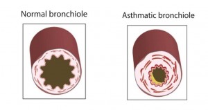
Asthma is chronic respiratory disorder with intermittent exacerbations that can be brought on by a variety of environmental, infectious, physical, and even emotional triggers.  The hallmarks of asthma are varying degrees of difficulty breathing along with wheezing.  Wheezing is a respiratory noise made typically with expiration and is a result of narrowing of the “middle” airways. Three pathophysiologic processes lead to this narrowing of airways: bronchospasm, inflammation, and excess secretions (fig 10).  The result of these abnormal processes is decreased air exchange with the alveoli, which leads to a decrease in O2 and an increase in CO2 in the bloodstream.

Figure10-resp

Figure 10 Notice that in asthma the muscle layer of the bronchiole hypertrophies (thickens), the lining of the airway becomes edematous (swollen), and secretions (excess secretions from the lungs and/or pus from infection) fill the lumen of the airway. All of these processes result in narrowing of the airway


Patients with asthma have a tendency to develop this response to a variety of stimuli: allergies, respiratory infections, emotional stress, to name a few.  The individual with asthma may have perfectly normal lung function until a reaction is triggered by allergens such as mold or pollens, irritants such as smoke, or virus infections of the respiratory tract. In some cases, no external stimulus can be identified as the cause of an asthma attack.
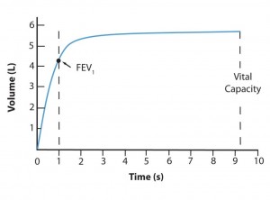
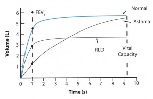
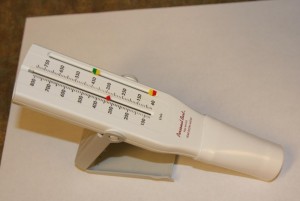
While the impairment of gas exchange is reflected by hypoxia and hypercarbia, a more graphic picture of the mechanical aspect of asthma can be found in the alterations of mechanical parameters found on pulmonary function testing.  There are a variety of pulmonary function tests available for this, but the simplest and most useful measurements are: 1) FEV1 (Forced Expiratory Volume one), the maximum amount of air that can be expired by a patient in one second of forceful exhalation (Figures 11 and 12); and 2) Peak Expiratory Flow Rate (PEFR), which represents a “snapshot” of fastest rate of airflow the patient can achieve during this forceful expiration.  In asthma and other bronchospastic conditions, both the FEV1 and the peak expiratory flow rate are diminished. The PEFR is easiest to measure (in liters/min), with a simple hand-held device (Figure 13).

image13

Figure 11 A normal pulmonary function curve plotting forced expired air volume against time in seconds. In this example of a normal adult, the FEV1 is about 4.4 liters and the Vital Capacity (VC, the total functional lung volume), is about 5.8 L. FEV1% is the percent of air expired in 1 second compared with all of the air expired: in other words, FEV1% = FEV1/VC (100). A normal FEV1% is about 75%.

image14

Figure 12 Another set of pulmonary function curves comparing normal with asthma and restrictive lung disease (characterized by decreased lung volume due to a variety of causes). Note that the asthmatic curve eventually approaches a normal vital capacity, but rises much more slowly than normal. The curve for RLD rises in a fairly normal fashion, but levels off at a lower vital capacity.

image15

Figure 13 Peak Expiratory Flow Meter. The Patient blows into the mouthpiece (on the right) as hard as possible and the maximum rate of air flow is measured by the little red arrow in the middle.

As with most respiratory problems, the patient’s main complaint is that of difficulty breathing or dyspnea (a feeling of shortness of breath).  Because asthma tends to be a recurrent disease, eliciting a history of previous asthma attacks can be helpful.

On physical exam, the patient’s respiratory rate is usually increased in an attempt to compensate for the inefficiency of each breath.  There is visible effort required to achieve inspiration and expiration.  The nostrils may dilate and there may be use of neck muscles to assist with inspiration.  Listening to the lungs with a stethoscope may reveal wheezing, a cluster of high-pitched whistling sounds reflecting partial obstruction of multiple small airways (bronchioles).

If the level of oxygen in the blood is low enough, the patient’s skin, lips, and nail beds may be cyanotic (literally, “blue” reflecting the predominance of de-oxygenated hemoglobin). One way of quantifying this is the bedside measurement of oxygen saturation (SaO2).  This technically simple process involves attaching a small painless probe to some area of the body such as a fingertip or earlobe.  A tiny beam of light passes from one side of the probe through the tissues to a receptor on the other side.  Because oxygenated hemoglobin (Hgb) and de-oxygenated Hgb absorb light differently, the degree of saturation (the proportion of the blood’s hemoglobin that is bound to O2 vs. that bound to CO2) can be measured.  This “pulse ox” measurement reflects the ability of the respiratory system to provide this O2 to the bloodstream.  At sea level, close to 100% of Hgb should be saturated (a SaO2 of 100%).  A SaO2 of less than that requires explanation: less than 90% often requires intervention, and sudden drops below 80% can result in loss of consciousness.  Oxygen saturations below 50% reduce the supply of O2 to the heart enough to result in cardiac arrest and death.

A more complete picture of the O2, CO2, and acid-base status of the blood can be determined by drawing blood for blood gasses. This test is usually drawn from an artery (arterial blood gas or ABG), and renders values of the blood pH (measure of acidity), and the partial pressures (remember Torricelli) of oxygen (pO2) and carbon dioxide (pCO2).  Normal values for an arterial blood gas are:

pH: 7.35 –7.45

PaCO2: 40 mm Hg

PaO2:  80 – 100 mmHg

Imaging of the anatomical components of the respiratory system is another important diagnostic modality. A chest x-ray provides a two-dimensional “shadow” picture of the internal structures of the chest. While it may not provide as much anatomic detail as other imaging modalities (e.g. computerized tomography, magnetic resonance imaging, etc.), a chest x-ray is relatively cheap, quick, and painless, and results in very low level of radiation exposure.  A long history of experience with x-rays of the chest has shown that this study is very good at picking up conditions that can complicate asthma management: e.g. pneumonia, pneumothorax, and atelectasis.

As stated in the beginning of this book, physicians treat disease by directing therapies at the underlying pathophysiologic process.  Let’s look again at the pathophysiology and consider the interventions that may help in asthma.

The wheezing associated with asthma is a reflection of airway narrowing, at least in part due to bronchospasm.  To relieve bronchospasm we need to use a medication that will relax airway smooth muscle (so it doesn’t contract so much and constrict the airway).  Medicines that relax airway smooth muscle are called bronchodilators.  The most frequently used bronchodilator is albuterol, a drug of the beta-adrenergic class. Anticholinergic drugs (substances that block the bronchoconstricting and secretory effects of the neurotransmitter acetylcholine) are also useful adjuncts in acute exacerbations of asthma.  These drugs are weak bronchodilators compared to “beta 2 agonists” like albuterol, but work to dilate the airways by a different mechanism. Though not typically used as “first line” agents for asthma, they are often used is severe asthma exacerbations as an adjunctive therapy. Examples of these anticholinergic agents are atropine and ipratropium. Epinephrine (the active agent in many over-the-counter inhalers) is another great bronchodilator but it has many cardiac effects even when inhaled (causing the heart to beat faster and work harder), so it is a less desirable agent.  All of these agents are typically aerosolized and inhaled by the patient so the medicines can be delivered directly to where they are needed thus minimizing untoward effects elsewhere in the body.  Bronchodilators can be used on a regular basis to keep the disease in check or intermittently to treat exacerbations.

Because of the inflammatory component of this disease, anti-inflammatory drugs are frequently used to treat acute asthma.  While there are several categories of anti-inflammatory agents, the most commonly used are the adrenal corticosteroids (similar to the steroids produced by our own adrenal glands).  These agents serve to decrease inflammation of the airways, thus increasing their internal diameter.  These drugs can be administered orally, by inhalation, or by injection. While these medications have little effect immediately, they make dramatic changes several hours after the start of an attack and are highly effective in long-term disease control and eventual termination of an acute asthma attack.

Accumulation of fluid in the airway is the third and final pathophysiologic process that needs to be addressed.  Airway secretions are often increased in the setting of allergic reactions and other asthma triggers. Mucus accumulation in the airway can lead to plugging and lack of airflow to the lower airways and alveoli. Current asthma treatments do little to decrease mucous secretions in the bronchial tubes, but novel medical therapies are in the process of being developed. In the meantime, we can only suction and lavage (wash out or “irrigate”) the airways to clear this obstruction: for those patients sick enough to be on a ventilator. Infection can be another source of increased airway secretions. While many of these infections are caused by viruses (for which there is typically no treatment), infectious secretions can sometimes be produced by bacteria that grow in the airway and cause the production of pus. This fluid, too, will lead to airway narrowing and obstruction to air flow. Antibiotics are used to eradicate the pus-producing bacteria.  Bacteria and pus formation also induce inflammation and trigger bronchospasm so it is very important that this be treated early.

Removing triggers of acute asthma (certain foods or environmental allergens) is also another very important part of the care of any asthma patient. Using immunotherapy and allergy medications may also help to prevent an asthma attack in patients sensitive to certain allergens, e.g. ragweed, or bee stings. In someone already experiencing a flare up of their asthma there are a variety of supportive measures that are often required.  If a patient is hypoxic, supplemental oxygen may be given for the patient to breathe until the therapies above can be instituted. In patients with profound difficulty in breathing, you simply follow the ABC’s: airway, breathing, and circulation.

Many patients with an acute asthma attack will require multiple treatments, directed at multiple pathophysiologic processes.  Most asthmatics with acute exacerbations will improve sufficiently during an emergency visit that they will get to the point that they will be able to be managed at home.  Most of these patients will require daily aggressive management, at least in the short term, to keep them from deteriorating again.  People do die from asthma attacks, so the medical team must be ready to intervene should the seriously ill patient require airway intervention in the event of a respiratory arrest.  An indication of impending respiratory failure can be a decrease in level of consciousness (sleepiness, difficult to arouse), inability to say more than a word or two between breaths, gasping with little air movement, struggling to breathe with severe diaphoresis (sweating).  These patients need to be treated rapidly and aggressively because they cannot keep up this kind of work of breathing for very long before they become utterly exhausted. At times the use of pressurized air by means of CPAP or BiPAP can assist the patient’s respirations and decrease the work of breathing.

Recalling the ABC’s, the most basic intervention for patients in acute respiratory arrest is the establishment of an open airway.  Depending upon the circumstances, this might be accomplished simply by properly positioning the patient’s head and neck and lifting the tongue off the back of the throat.  Some situations may require use of a suction device to clear the airway of secretions and/or vomit.  Others may require that the physician place an artificial airway (an endotracheal tube) into the trachea (endotracheal intubation).  If breathing is inadequate in rate or depth, it must be assisted by some means.  These include mouth to mouth ventilation, use of a facemask attached to a resuscitation bag that delivers air when compressed, or a mechanical ventilator (or respirator) that is attached to the endotracheal tube and controls air delivered to the lungs.  These measures will need to be continued until the patient’s respiratory system recovers to a point that he or she can again breathe on their own.

Anatomy & Physiology

As stated above, the respiratory system is an interface between our body and the environment. Just as the digestive system exchanges nutritional substances between the environment (“the outside”) and the bloodstream (“the inside”), the respiratory system is the mechanism by which gasses move into and out of our internal environment. The gasses exchanged by the respiratory system play a key role in the metabolism of nutrients brought into the body by the digestive system.

Oxygen (O2) is the most important gas entering the system. It is one of the necessary substrates for cellular energy production. This is sometimes referred to as cellular respiration. Since we cannot store oxygen to any significant degree, even brief disruptions in oxygen intake from the environment can be devastating. The most important gas exiting the respiratory system is carbon dioxide (CO2). Carbon dioxide is a cellular waste product. Continuous removal of CO2 is vital to normal cellular function. Accumulation of CO2 in the blood and tissues results in alteration of the acid-base status of the body. Progressive excess of CO2 leads to sedation, stupor, coma, and ultimately death.

The respiratory system, like the cardiovascular system, must function continually. For this reason, in virtually all medical emergencies, support of respiratory function is of the greatest importance. This principle is often referred to as doing the ABC’s which stands for Airway, Breathing, and Circulation. Without an open passage for air exchange, all other measures will be futile; the same applies to breathing (also referred to as ventilation). Thus, one could argue that the respiratory system is our most important system since all resuscitations start here.

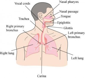
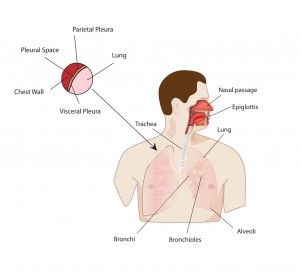
The respiratory system is sometimes referred to as the “respiratory tree” because of the way it branches like an inverted tree into smaller and smaller airways before getting to the alveoli where gas is exchanged. To simply categorize this “tree,” it can be separated into 4 levels (Figure 1).

  1. The Oropharynx and Nasopharynx
  2. The Larynx and Trachea
  3. The Bronchi and Bronchioles
  4. The Terminal Bronchioles and Alveoli

The respiratory system begins at the nose and mouth: both are portals that define the start and finish of air exchange. Of the two portals of entry, the nasal passage is narrower and offers more resistance to airflow, but has a moist mucosal lining that does a better job of filtering and humidifying incoming air. The oral passage allows a greater rate of air movement with less resistance, but filters and humidifies air less effectively. Both passages join in the posterior pharynx and inhaled gas then goes through the opening (the glottis) at the top of the larynx, and then into the trachea.

The larynx, housing the vocal cords, has the narrowest aperture of these structures. Edema (swelling) at this level can greatly impact air flow. Foreign bodies commonly impact and obstruct the airway at this level. Muscles in the larynx can control the aperture at this level as well, and spasm of these muscles can lead to impaired air intake. Beyond the larynx, the trachea is little more than a rigid tube that divides into right and left mainstem (or primary) bronchi, each leading into a branching system of ever-smaller airways.

image05

Figure 1 Anatomy of the respiratory system: overview.

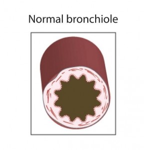
Bronchi and bronchioles are more functionally dynamic than the trachea. Their walls have a thick muscular layer that contracts and dilates to change the diameter of the airway (Figure 2).

image06

Figure 2 Drawing of a bronchiole in cross-section. The walls of the bronchiole contain smooth muscle, which contracts and relaxes to alter the size of the air passages.

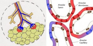
This branching system of airways ultimately terminates in the alveoli: tiny sacs with walls just one cell layer thick (Figure 3). This thin membrane is all that separates the gas-filled alveolus from the blood-filled capillaries; through this membrane, oxygen and carbon dioxide pass between the air and the bloodstream.

figure3-resp

Figure 3 Left: bronchioles terminating in alveoli, schematic drawing. Right: detail showing blood flow to the individual alveoli. Deoxygenated blood (represented by a blue color, even though this blood is actually dark red and not blue) enters the alveolar system, acquires oxygen, and exits as oxygenated blood (red portion of vessels). What are not shown are the small terminal bronchioles that attach to each of the alveoli.

In addition to familiarity with the internal structures of the airway and lung, knowledge of the anatomy of the chest is also essential for understanding the process of respiration. The chest is structured around a bony cage formed by the ribs, which are attached to the sternum, in front, and the spine posteriorly. The attachments are such as to allow a certain amount of motion of the ribs with respect to the spine, sternum, and to one another. The ribs are interconnected by muscle; external to this rib-muscle layer is more muscle, and external to this is subcutaneous tissue and skin. Internal to the rib-muscle layer is a tough, thin, leathery membrane known as the pleura, which forms the lining of the chest cavity. (We talk about the chest cavity as if it were a single space, but functionally there are two spaces, right and left, separated by the mediastinum which contains the heart, trachea, esophagus and other important structures.)

image09

Figure 4 Upper Left: detail of the interface of the lung with the pleura and chest wall.

Following the pleura into the recesses of the chest cavity, we find that it folds back upon itself to form an envelope: the pleural space (Figure 4). We have already seen that one surface of this envelope, the parietal pleura, is attached to the inner chest wall. The other surface covers and adheres to the outer surface of the lung: the visceral pleura. Between the two pleural surfaces is a potential space with a negative air pressure (a vacuum). This vacuum must be present in order for breathing to function normally: part of the mechanics of breathing, which we will cover next.

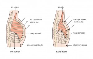
To move air into the lungs, the muscles of the chest wall contract, expanding the ribcage; the diaphragm contracts as well and lowers the floor of the chest cavity. The net effect is to enlarge the chest cavity (Figure 5). Because the parietal pleura is attached to the inside of the ribcage, it too expands outward. The vacuum in the pleural space ensures that the visceral pleura expands as well and brings with it the underlying lung to which it adheres. Expansion of the lung creates the negative pressure that sucks air in through the airway, completing the process of inhalation (also called “inspiration).

image01

Figure 5 The mechanics of breathing. Left: inhalation (inspiration), showing expansion of the chest cavity. This increases the volume of the chest cavity and lungs, creating the negative pressure that draws air into the airway (see text for details). Right: exhalation (expiration), a passive process.

Exhalation (known medically as expiration) is not as complicated. When the chest muscles relax, the chest recoils to its resting size, and air moves passively out through the airway. It is important to note, however, that the lungs do not expel all of the air they hold. The leftover air, after full exhalation, in the lower airways and alveoli (which do not collapse) is termed the residual volume. In adults, that volume is about 1 liter. Thus, the air taken into the depths of the alveoli with the next breath is not entirely the same as atmospheric air but is a mixture of environmental air and some of the air that was left from the previous breath. This is an important concept in that the partial pressure of retained carbon dioxide in the alveoli is in equilibrium with the major buffer in the body that determines the pH of the blood: bicarbonate. We will discuss this in much greater detail in the next chapter: The Renal System.

Introduction & Featured Case Scenario

The Respiratory System

We live in an ocean of air.*

Gabrielle Walker

Introduction:

The air in which we live is a mixture of gasses: about 21% oxygen, 78% nitrogen, and trace amounts of carbon dioxide and other gasses. Just as combustion requires a spark, fuel, and air, the human body requires oxygen in order to process nutritional substrates for energy and growth. These same processes produce carbon dioxide as a byproduct, and if allowed to accumulate it becomes toxic. The primary function of the respiratory system is the exchange of oxygen and carbon dioxide between the external environment and the bloodstream.

In this chapter, we will care for a patient with a serious respiratory problem. We will first learn about the anatomy, physiology, and pathophysiology of the respiratory system, and then highlight some of the clinical tools at our disposal to help in the diagnosis and treatment of respiratory ailments. Finally, armed with this knowledge, we will return to our patient and see what we can do to help him.

Case Scenario

You are a paramedic responding to a 911 call. At a nearby middle school, a 12 year old boy is having difficulty breathing. You arrive at the grade school with your paramedic colleague and are quickly directed by the principal to the school cafeteria. Across the room, sitting upright on a folding chair, you find a young boy struggling to breathe. His lips are blue, he is clearly frightened, and he is too breathless to speak. What do you do?

Take a moment to jot down some ideas on what could be wrong with this boy (i.e. think about pathophysiology) and what you could do to help him.

Natural Science Module Resp

The Respiratory System

We live in an ocean of air.*

Gabrielle Walker

Introduction:

The air in which we live is a mixture of gasses: about 21% oxygen, 78% nitrogen, and trace amounts of carbon dioxide and other gasses. Just as combustion requires a spark, fuel, and air, the human body requires oxygen in order to process nutritional substrates for energy and growth. These same processes produce carbon dioxide as a byproduct, and if allowed to accumulate it becomes toxic. The primary function of the respiratory system is the exchange of oxygen and carbon dioxide between the external environment and the bloodstream.

In this chapter, we will care for a patient with a serious respiratory problem. We will first learn about the anatomy, physiology, and pathophysiology of the respiratory system, and then highlight some of the clinical tools at our disposal to help in the diagnosis and treatment of respiratory ailments. Finally, armed with this knowledge, we will return to our patient and see what we can do to help him.