Exploring Medicine – A Foreword

I remember clearly the first meeting I had with Steve Jameson in summer 2005. I had just returned from a regional meeting of the National Association of Advisors in the Health Professions (NAAHP) where a number of sessions had focused on the importance of having health professions advisors build partnerships with health care practitioners in the community. So, when approached by Steve I was more than glad to meet to discuss what I thought was merely an opportunity to enhance our students’ access to shadowing opportunities.

Although that original meeting has led to much greater student access to clinical experiences, then I had no idea that it was the launching pad for an innovative educational experiment in the health sciences that has deeply contributed to the academic, professional, and personal development of hundreds of students – Exploring Medicine. In our first conversations in 2005, Steve proposed to teach a course at the College of Saint Benedict/Saint John’s University (CSB/SJU) that would show students how to critically think like a doctor and how to apply the material they were learning in basic science courses to the process of clinical diagnosis.

We eagerly supported Steve’s idea and in spring 2006 Exploring Medicine was taught for the first time. Since then it has been taught every spring at CSB/SJU and more recently in the fall semester at the University of St. Thomas. Exploring Medicine is truly a gem since it allows students to critically engage in a focused and structured learning experience from a clinical standpoint. Through Steve’s interactive lectures students develop analytical skills that allow them to experience the thinking process of a clinician making a diagnosis; through the panels of healthcare professionals and guest speakers students gain an appreciation for the diversity of the healthcare field and the necessity for a team-approach in care; and last but not least, through the structured shadowing experiences provided by the class students can see how principles covered in the course are directly applied in the process of diagnosis and patient care. Indeed so many students continue to pursue the relationships they have established with clinicians in Exploring Medicine that at CSB/SJU, with Steve Jameson’s help, we established a year-long internship program at the St. Cloud Hospital Emergency Department entitled the Student Health Assistant Program. Engaging community physicians to teach at local universities pays dividends, and Exploring Medicine is the ideal platform to establish those relationships.

The uniqueness of Exploring Medicine is that it is not static – Steve Jameson is continuously editing and tailoring his presentations, creating new experiences for students, and providing novel leaning tools and settings. Since 2006 Exploring Medicine also came to include this book – it is used for the class, but many Exploring Medicine alums will also vouch for its continued value as a refresher and review tool in their further studies. More recently Steve has developed online resources that allow Exploring Medicine to be delivered in its unique and creative fashion to health professions students in any college campus throughout the country; tools that were recognized by the AAMC with its 2013 iCollaborative award in biology.

In short, Exploring Medicine is much more than a course, a book, or a set of online tools. Exploring Medicine is a unique experience that allows health professions students to directly bridge their academic background to a structured clinical setting, and to begin to experience the intellectual world as seen through the eyes of a physician. In the process of helping Steve implement his vision at CSB/SJU, I have seen hundreds of students who are now working as physicians, physician assistants, physical therapists, among others engage in their first meaningful clinical discovery in that setting. Exploring Medicine is indeed the bridge from the world of the humanities, social and natural sciences of our college campuses to the experiential setting of clinical medicine and practice.

Manuel Campos, Ph. D.

Professor of Biology, Preprofessional Health Advisor College of Saint Benedict|Saint John’s University

Preface

Choosing a career is one of the most important decisions you will make in your life. While a career in health care, and in medicine in particular, can be incredibly exciting and rewarding, the journey to that end can be an enormous physical, emotional, and financial drain. The decision to go into this field must be an informed one.

Many are enamored with the medical field and with “being a doctor” long before they know much at all about the practice of medicine. Some are told, “You’re smart; you should be a doctor.” Others simply like what they see on TV. The Exploring Medicine series of modules will primarily focus on what it is like to be a physician, but the information is relevant to all that are seeking a career as a health care professional. To make an informed decision regarding a career in medicine, you should first explore the medical field by seeing what physicians do and learn to think like a physician.

This module, and others in this series, will allow you to do just that. Starting with the very first topic, you will plunge into the world of clinical practice. There you will find patients with a variety of medical problems (based on actual cases), many with life-threatening and life changing emergencies that you will need to work through and solve in order to save your patient’s life and make them well. By the time you have finished this module you will have learned to think and problem-solve like a doctor, and you will understand the process of diagnosing and treating disease.

The medical model is all about diagnosis and treatment of disease, and in order to understand that you have to understand pathophysiology. Pathophysiology is the study of disease. The pathophysiologic process of a disease is the series of events that take place and conditions that develop that result in a particular disease. In order to treat disease, the physician must understand something about the pathophysiologic process that led to the development of that disease. By going through the systems based modules you will gain insight into the thought process physicians use to diagnose and treat disease, and how they help patients to become well. Each system based module begins with a general overview followed by a discussion of the pathophysiology of a particular disorder: e.g. the Respiratory module reviews anatomy and physiology of the Respiratory System, then goes into a discussion regarding the cause of asthma and its treatment. Other modules, like Social Determinants of Health, focus on wellness as it relates to society, social factors, and public policy. Exploring Medicine modules were made for the students that have taken biology, chemistry, sociology, psychology, and other science courses, and wondered, “Why do I have to learn this stuff?” These modules apply the student’s science knowledge to the real world by making correlates to clinical medicine.

Books have been written on what will be small elements of the Exploring Medicine modules, so we obviously cannot comprehensively cover every topic. The authors have distilled a massive volume of material down to several manageable lessons. Despite this editing, the amount of subject matter may still seem intimidating at first, but that is not the intent of the authors. Do not get stressed or anxious. The focus of these modules will be on general principles and concepts. In order to transition from simple anatomy and physiology to treatment of disease, for example, we must streamline our approach in order for the reader to get some appreciation of what it is like to be a physician. The following module will take you, the medical enthusiast, through a very narrow slice of medical training: from medical school, to residency training, to clinical practice. Armed with your newly found knowledge, you will be able to make clinical decisions based on true-to- life patient scenarios and help your patients become well or stay well. If you find this process compelling, as the authors do, then you may seriously want to pursue the exciting field of medicine.

Introduction to the Chapters – Life and Death, Homeostasis and Equilibrium

Death is not the enemy but occasionally needs help with timing. – Peter Safar, M.D. The “Father” of CPR

What is life? That may seem like a metaphysical question, but it’s actually not. It is a very real question for those that provide medical care, but the answer can be surprisingly complicated. As a physician, physician assistant, nurse, or nurse practitioner, you will likely have many occasions where you will be dealing with patients as they look to cross that line from life to death. In the following chapters we will discuss a variety of pathophysiologic processes that will alter the normal function of the human body.

Before we delve into specific disease processes, though, let’s take a look at a more fundamental principle of human physiology and how a disruption of normal processes can lead to chaos, i.e. entropy, on a microscopic (actually molecular) level and to death on a macroscopic level.

The human body is a complex network of systems that operate in unity to keep us alive and active. In order to maintain normal function the body seeks homeostasis and defies equilibrium. Cellular reactions take place under very specific conditions: a narrow range of pH, the correct concentrations of particular ions, proper amount of substrates, etc. Homeostasis is the set of conditions that the body establishes and maintains in order to function properly. It takes energy to maintain this homeostasis. This energy has to be consumed in the form of chemical bonds contained in the food we eat, converted to a usable form, transported throughout the body to all of its cells, and metabolized into a form of energy the cells can use: ATP. The wastes of these reactions then have to be removed. It is a complex network of integrated systems that accomplishes all of these tasks.

In Exploring Medicine, we will look at four of the body’s integrally related physiologic systems (Cardiovascular, Respiratory, Renal and Integumentary) as examples of how the body maintains homeostasis. We also explore what can happen when these systems fail, and what we, as medical care providers, do to restore normal function and reverse the process of dying. All of the chapters have questions to answer along the way and challenging scenarios to solve at the end of each chapter. Answers to these questions and further elaboration of concepts are in the appendices near the end of the book. In the chapter on Evidence Based Medicine, we will discover how the medical literature helps to guide a physician’s clinical practice. As you strive to learn more about the field of medicine, remember the words of Hippocrates (the final line of the Hippocratic Oath), “May I always act so as to preserve the finest traditions of my calling and may I long experience the joy of healing those who seek my help.”

Renal System Unit Quiz

This is an open book test.  You can click here to open the lesson in a new tab/window to review the lesson as you take the quiz.  If you close the quiz you will have to start all over with a different set of questions. You need to achieve a score of 90% or greater in order to obtain the certificate of completion.

Please ensure you have enough time to complete the entire quiz at one time.

Acid/Base Pathophysiology

There are 4 basic acid/base disorders: 2 metabolic disorders (metabolic acidosis and alkalosis) and 2 respiratory disorders (respiratory acidosis and alkalosis). These disorders can, for the most part, be explained by the using the carbonic acid equation:

image01

Disorders that move the carbonic acid equation to the left, or result in greater H+ concentrations, cause acidosis. The converse is true, in that disorders that move the equation to the right, or result in a lesser H+ concentration, cause alkalosis. There are two exceptions to this rule: 1) Drinking lots of water (adding substrate to the right side of the equation) does not result in any significant acidosis, and 2) A disorder resulting in increased H+ will move the equation to the right and HCO3will be used up, but there is still an increase in H+ so there is acidosis, not alkalosis. Each disorder will be discussed individually.

Respiratory acidosis:

Respiratory acidosis results from an excess of CO2 in the serum. This typically occurs because normal air exchange is impaired at the level of the alveoli. Causes may be that the patient’s ventilations are suppressed, as can be the case with an opiate overdose, or that there is airway obstruction impeding adequate flow of air in and out of the alveoli, as can be the case with asthma or emphysema. The end result is a rise in pCO2 in the alveolus, which leads to a rise in serum pCO2, and this condition results in an increase in [H+] and decrease in pH.

Respiratory Alkalosis:

Respiratory alkalosis results from a decrease in CO2 in the serum. This typically occurs as a result of hyperventilation and the resulting increase gas exchange with the environment. pCO2 in the alveolus promptly drops to less than 40 mmHg, and then rapidly equilibrates with the alveolar capillaries and draws increased CO2 from the serum causing the serum pCO2 to go down. The carbonic acid equation moves to the right, as H+ binds with HCO3, and alkalosis ensues. Conditions that can cause this are reactions to certain drugs, anxiety, and certain metabolic states (to be discussed later).

Metabolic Acidosis:

Metabolic acidosis is the most complex acid/base disorder because there are so many causes for this condition. This is also an important topic because it is a common disorder and can occur in many different situations. In short, anytime there is a condition that results in excess acid production, increased H+ reabsorption (from the kidneys), decreased HCO3 production, or increased utilization or excretion (from the kidneys) of HCO3, the result is a metabolic acidosis. Because there are so many different causes of metabolic acidosis, we must break them into categories of anion gap and non-anion gap etiologies.

Anion Gap:

An “anion gap” (AG) implies that there is an anion (negatively charged particle) in the serum that is not accounted for. Basic chemistry tells us that a solution must exhibit electrical neutrality. That is to say that the number of charges of cations in the serum must be equivalent to the number of charges of the anions. Since the primary cation in serum is Na+(sodium), and the primary anions are Cl and HCO3, an equation can be written to show a balance between these charged particles:

AG = [Na+] – [Cl] – [HCO3]

K+ can be added to this equation as well, but it is usually ignored since its concentration does not change appreciably, even in extreme cases, and its concentration is relatively small. Under normal circumstances, the [Na+] is about 140 mmol/L, the [Cl] is about 105 mmol/L, and the [HCO3 ] is about 25 mmol/L. In this case, the anion gap would be 10. A normal AG is anywhere from 8 – 16 mmol/L (or more appropriately, meq/L). Given the example then, this means that, under normal conditions, proteins and other trace anions in the serum account for 8 – 16 meq/L of unmeasured, negatively charged particles.

Osmolal Gaps:

While we’re on the topic of measured and unmeasured particles, there is just one more concept to introduce: the osmolal gap (OG). Particles that are small enough and numerous enough to change the osmolality of the serum, but may or may not carry a charge, are considered osmotically active. The major osmotically active particles in serum are sodium and its associated anions, glucose, and urea (a waste product of protein metabolism). When urea is being carried in the blood, it is called “blood urea nitrogen” or BUN. As you may have guessed, there is an equation to calculate serum osmolality:

2[Na+] + [glucose]/18 + [BUN]/2.8

Sodium concentration, measured in meq/L, is multiplied by 2 in order to account for the anions (chloride, bicarb, proteins, etc.) associated with it. Glucose and BUN concentrations are measured in mg/dL, and are divided by 18 and 2.8, respectively, to account for their osmotic activity relative to their molecular weights. The normal calculated osmolality of the serum is 280 – 300 mosm/kg. Serum osmolality can also be measured, typically by means of determining freezing point depression. There are, of course, other osmotically active particles in serum that are not measured by the formula above. The difference between the measured serum osmolality and the calculated serum osmolality is called the osmolal gap (OG):

OG = serum osmmeas – osmcalc

A normal serum OG is less than 12. If an individual ingests a substance that results in increased osmotically active particles in the serum, then there will be an osmolal gap that is greater than normal.

Non-Anion Gap Metabolic Acidosis:

Non-anion gap metabolic acidosis results from a loss of bicarb. The other major anion in serum, Cl-, goes up in this condition (to maintain neutrality of charges in the serum) and thus this condition is also called hyperchloremic metabolic acidosis. Bicarb is a measured particle and thus there is no anion gap when the AG is calculated with this acidotic state. Medical conditions that can cause a non-anion gap metabolic acidosis are:

  1. Diarrhea
  2. Certain drug ingestions (e.g. acetazolamide, which interferes with HCO3formation)
  3. Renal dysfunction

Anion Gap Metabolic Acidosis:

The most complicated category of acid-base disorders is that of anion gap metabolic acidosis (AGMA). For those who like to play the part of Sherlock Holmes and solve mysteries or, more contemporarily, play the lead detective in CSI (crime scene investigator), this is the category for you. Many conditions or ingestions of toxins can result in an anion gap metabolic acidosis. In order to determine the cause of this acidotic state, we have to categorize these patients based on the patient’s history and physical exam, and on lab tests: e.g. determine if there is an osmolal gap.

Many substances and conditions can cause an anion gap metabolic acidosis. A useful, but not quite complete, mnemonic is MUDPILES. This stands for:

Methanol – turns to formic acid

Uremia acidosis from renal failure

Diabetic ketoacidosis – develop ketoacids” in Type I diabetes mellitus

Paraldihyde – a drug rarely used, develop lactic acid

Isoniazid, Iron – excess of both result in cellular anaerobic metabolism and lactic acid formation

Lactic acid – many causes of anaerobic cellular metabolism

Ethylene glycol – metabolized to glycolic acid

Salicylates – salicylic acid and causes anaerobic cellular metabolism and formation of lactic acid

All of these substances can cause an anion gap metabolic acidosis. For a list of those that cause and do not cause an osmolal gap in the setting of this acidosis, refer to the list below.

Examples of Conditions and Substances that Cause AGMA With Normal and Elevated Osmolal Gap

Normal Osomolal Gap                                                         Elevated Osmolal Gap

1. Diabetic Ketoacidosis                                                                                          1. Methanol

2. Lactate                                                                                                              2. Ethylene Glycol

3. Salicylates                                                                                                         3. Alcoholic ketoacidosis

4. Toluene

Compensatory States for AGMA:

It is not uncommon for the pH to be profoundly low in a state of an anion gap metabolic acidosis. In order to compensate for this condition, the respiratory system is triggered to breath more rapidly and more deeply in order to exchange a greater volume of air with the environment and lower the pCO2 in the alveolus. This increase in respirations ultimately leads to a decrease in pCO2 in the serum, and an increase in pH. A compensatory response will only bring the pH up to a point that it gets close to a lower normal range; compensatory excess respirations will not be such that the pH will ever become greater than 7.40 (alkalotic).

Metabolic Alkalosis:

The last acid/base condition to discuss is metabolic alkalosis. This disorder typically results from loss of H+ and accumulation of excess HCO3 from either vomiting or because of a disturbance in normal renal function (often a drug effect). The accumulation of HCO3 can also be a compensatory effect in an effort to normalize serum pH in long-standing respiratory acidosis (e.g. in emphysema patients).

Review of Renal Case Scenario

You are the physician on duty in the ER when a middle aged male, whom you recognize from numerous previous visits related to alcohol intoxication, presents unresponsive. Your immediate response is to attribute this episode, as well, to alcohol intoxication. A friend that arrives with him, however, states that he has made sure that there is no booze in the home because he wants his friend to “dry out.” The patient is breathing rapidly, and upon further questioning the roommate informs you that there was an empty container of “camping fuel” lying on the floor next to the couch on which the patient was found.

On examination of the patient, you find the vital signs to be a blood pressure of 120/80, heart rate 130 beats/min, respiratory rate 30 breaths/min, and temperature of 98.6oF. He will only grimace to a deep painful stimulus (a strong knuckle rub to the sternum). You order some blood studies and get the following results:

serum ethanol level=.005g/dL of 5mg/dL

measured serum osm=318

image02

What is the acid base disorder? Show all of the necessary calculations. See the step by step solution to this case in the paragraphs that follow.

To be a good clinician in this challenging case you have to utilize some serious investigative skills: i.e. be like Sherlock Holmes. Since the patient’s pH is low, this is a condition of acidosis. Next, determine if the acidosis is secondary to a respiratory or metabolic condition. Note that the bicarb is low, so it is being used up by hydrogen ions, thus this is a metabolic acidosis. Note as well that the pCO2 is also low. This low CO2 is the result of excess respirations in an attempt, by the body, to at least partially reverse the acidosis, and thus there is appropriate respiratory compensation for this metabolic acidosis.

After a few calculations, you discover the following:

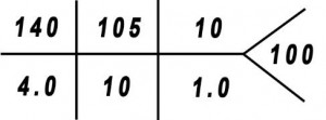
AG = 140 – 105 – 10 = 25

serum osmcalc = 2(140) + 90/18 + 9/2.8 = 288 taking EtOH into account add 5/4.6 = 1

so serum osmcalc = 289

osm gap = 318 – 289 = 29 (this is an elevated osmolal gap)

You determine that this is an anion gap metabolic acidosis with an osmolal gap. The differential diagnosis includes either poisoning from methanol or ethylene glycol, or alcoholic ketoacidosis. A screen for volatile substances in the blood is positive, so that means there is either methanol or ethylene glycol in the blood: so this is a poisoning. You direct your treatment at the underlying pathophysiologic process and order intravenous ethanol (explanation of treatment is below) and hemodialysis. A subsequent methanol level comes back high, and an ethylene glycol level comes back at zero: this confirms the diagnosis of methanol poisoning. The patient survives and has no long-term disability from this ingestion. He is successful in alcohol treatment and, with his friend’s help, remains sober.

Additional Information About Methanol Poisoning:

Methanol (CH3OH) is an alcohol that is found in many places: e.g. as a gasoline additive, windshield cleaner, fuel for camp stoves, “wood alcohol” as a byproduct of fermentation when attempting to make ethanol. Methanol can be physiologically harmful and even lethal in relatively small amounts.

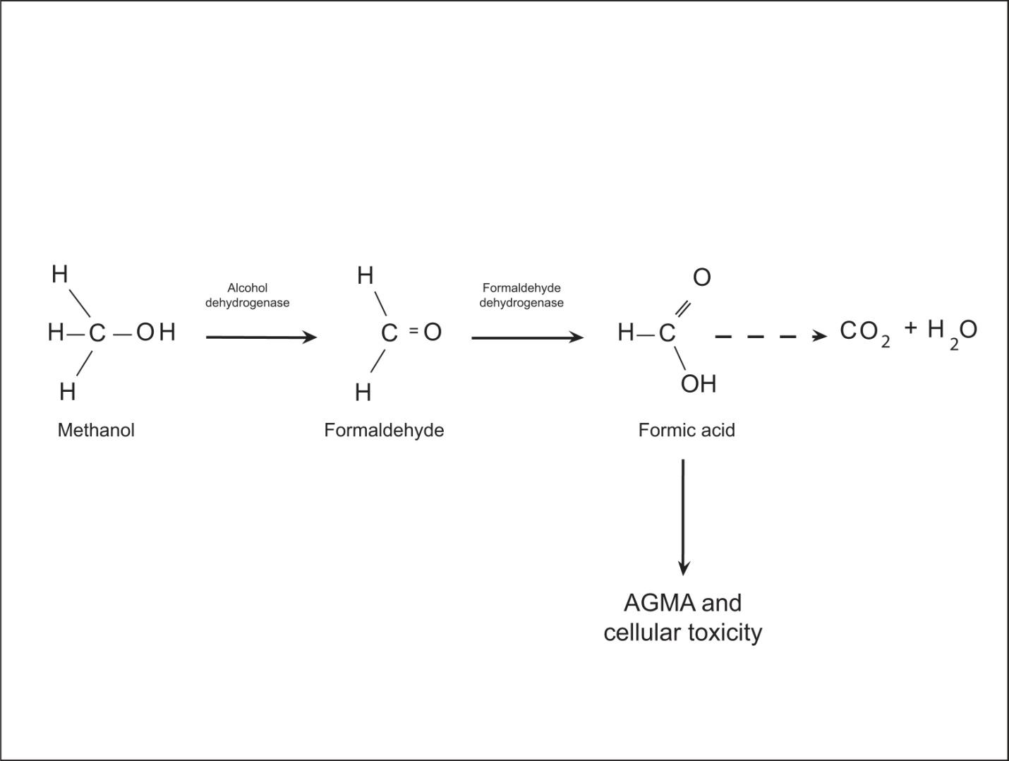
After ingestion, methanol is metabolized in the body by the same pathway that ethanol is metabolized (Figure 7): ethanol and methanol are both metabolized by the enzyme alcohol dehydrogenase.


image04

Figure 7 The breakdown (metabolism) of methanol in the body ultimately results in the formation of formic acid which is very toxic to human cells and can result in blindness, severe acidosis, and even death if not treated urgently.

Methanol, by itself, is osmotically active, but has little effect other than it can cause intoxication. It is, however, ultimately converted to formic acid: a potent acid that has devastating effects on tissues of the body. Most characteristically, even in small amounts, methanol can result in permanent blindness. If the metabolism of methanol can be slowed, then the body can manage limited amounts of formic acid and safely convert it to CO2 and H2O (Figure 8). The fastest and easiest way to slow methanol metabolism is to overwhelm the first step in the metabolic process: occupy the receptors of alcohol dehydrogenase. In order to accomplish this, the clinician induces a state of intoxication in the patient by administering ethanol (typically intravenously, but this can be performed orally as well). Fomepizole is a more contemporary (and much more expensive) agent used to competitively inhibit alcohol dehydrogenase and slow the metabolism of methanol. In severe cases, hemodialysis can be a useful adjunct as well.

image20

Figure 8 If allowed to metabolize normally, methanol quickly converts to formic acid and causes severe metabolic acidosis and cellular damage. If the conversion to formaldehyde is competitively inhibited by increasing the level of ethanol (or fomepizole) in the blood, the body can slowly metabolize the little bit of formic acid that comes through and minimize the toxic effect on the body.

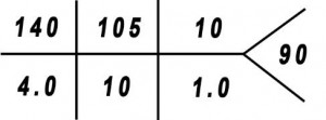
To diagnose methanol poisoning, the physician must first suspect the diagnosis, and then order the appropriate screening labs. Serum methanol levels cannot readily be obtained in most hospital laboratories (can take several hours to get results), thus the physician must use tests that indirectly indicate its presence. Routine lab studies would include a “chem 7,” ABG, and serum osmmeas. Let’s walk through the calculations of a patient with methanol poisoning again. The results of the lab work could look something like this:

image21 image22

serum osmmeas = 322

serum EtOH = 0.000

With a pH less than 7.35, we know the patient is acidotic. With a bicarb level that is very low (10 mmol/L), we know this is a metabolic acidosis. With a pCO2 that is low, we know that this patient is appropriately attempting to compensate for this metabolic acidosis by blowing off CO2 and moving the carbonic acid equation to the right (as illustrated previously) in attempt to normalize the pH. Calculating the anion gap we find it to be 25 (140 – 105 – 10) which is high, and thus this is an anion gap metabolic acidosis (AGMA) with respiratory compensation. Next, we want to determine if there is an osmolal gap (OG). We take the measured serum osmolality and subtract it by the calculated serum osmolality (osmcalc = 2(140) + 100/18 + 10/2.8, which is about 289. So:

OG = 322 – 289 = 33, which is exceedingly high.

Once again, we learned that there are only 3 things that can cause an AGMA with an elevated OG, and they are methanol poisoning, ethylene glycol poisoning, and alcoholic ketoacidosis (a disorder in which muscle is metabolized in order to generate cellular energy because alcoholics do not have adequate stored glucose.

Ketones” are produced as part of this process). A screen for ketones is negative and thus this case is a poisoning of either methanol or ethylene glycol (treatment is the same so it doesn’t matter which it is).

Conclusion:

Appropriately diagnosing acid/base disorders in critically ill patients is vital since the treatment given will hinge on this diagnosis. Determining the acid/base state of a critically ill patient can often help the clinician direct his/her treatment at the underlying pathophysiologic process of the disorder. Below are several more acid/base case scenarios. By doing these you will greatly improve your understanding of these various processes. The worksheet at the end of this chapter also helps you to work through another acid/base scenario in a step by step fashion.

Other Clinical Renal – Acid/Base Scenarios

Based on the lab values in each of the clinical scenarios below, determine the presence of an acid base disorder and write the type of disorder on the line next to the values. For metabolic acidosis, tell whether there is an anion gap, osmolal gap, and/or expected respiratory compensation. For respiratory conditions, tell if it is an acute or chronic disorder.

Background, Renal Anatomy & Physiology

Proper cellular function depends on chemical homeostasis: keeping the body’s fluid, electrolytes, and pH within a very narrow range. For example, blood serum pH normally runs about 7.4. Enzymes, hemoglobin molecules, cell receptors, and other proteins go through slight conformational changes (change shape slightly) and begin to malfunction when the pH varies too far from normal. A sustained pH of just +/- 0.25 from normal is incompatible with life, and a change of +/- 0.5, even for a short time, is almost certain death. The human body has a complex system to compensate for the presence of excesses and deficiencies of fluid, electrolytes, and hydrogen ions (the concentration of which affects pH). This compensatory and regulatory system involves several organ systems and a buffering system. We will discuss the highlights of this process.

The primary organ for directly controlling fluid and electrolyte balance is the kidney. The kidneys also, along with the pulmonary system (as discussed in the last chapter), play a major role in controlling pH. We will start this chapter with a general overview of renal anatomy and physiology (referring to structure and function of the kidney), then go on to a discussion of total body water and its distribution to different “spaces” within the body. A discussion of electrolytes and their normal values in the various body spaces is next, followed by a lesson on acid/base balance. We will conclude this chapter by tying all of this information together in a study of various pathophysiologic processes.

Renal Anatomy and Physiology:

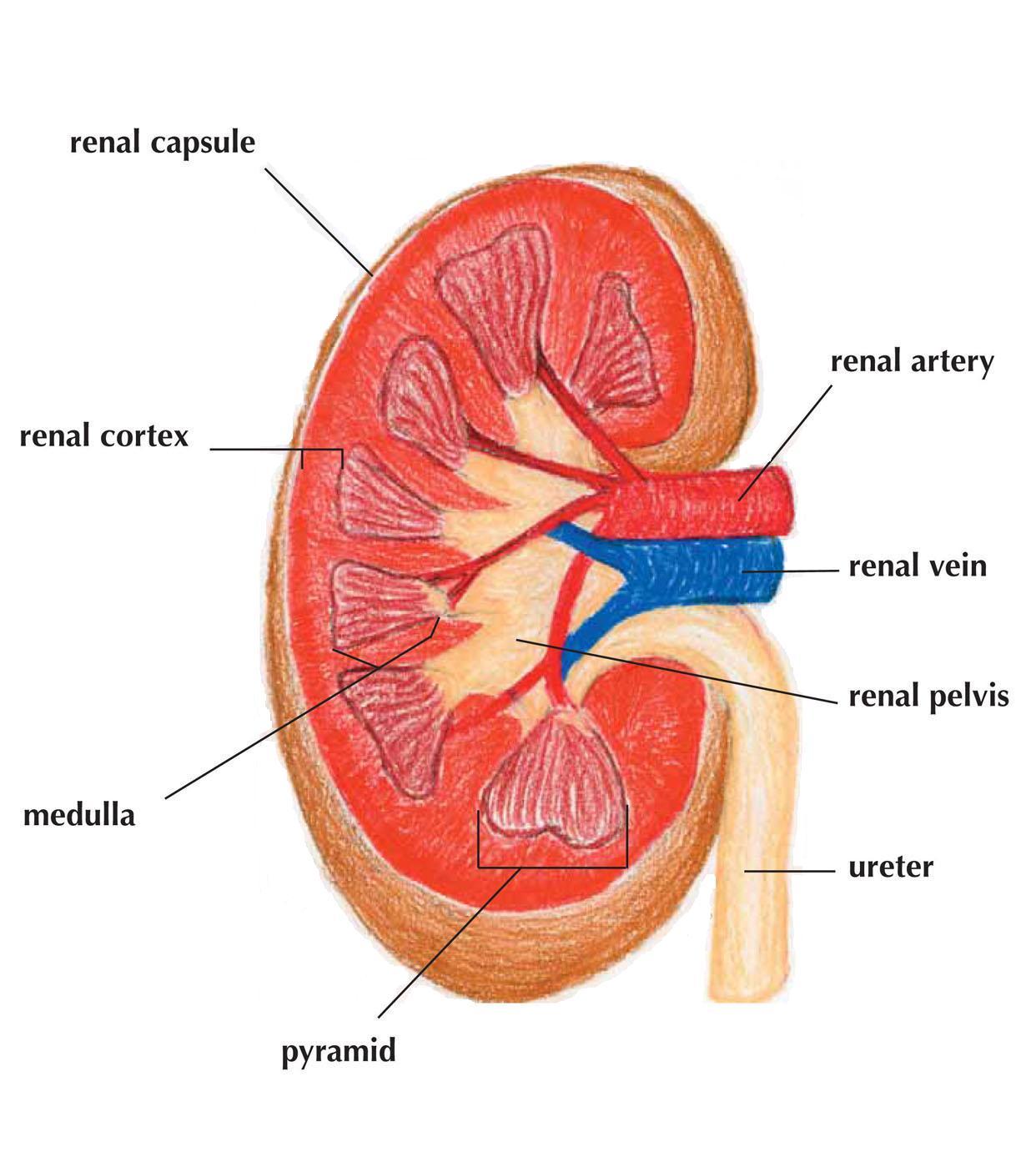
The kidney is made up of an outer portion, the renal cortex and medulla, and an inner portion, the renal calyces and pelvis (Figure 1). Within the cortex, and extending into the medulla, are millions of nephrons (Figure 2): blood filtering units that get rid of wastes, and regulate excretion and reabsorption of fluid and electrolytes. The waste ultimately becomes urine, and this is collected within the renal pelvis. It then travels through a tube (the ureter) to the urinary bladder, where it is ready to be expelled from the body. The kidney is the great regulator in the body in that it is capable of retaining more of a specific electrolyte when needed and getting rid of those that aren’t needed. When there are conditions of excess fluid (too much free water), the kidneys will expel hypotonic (dilute) urine. When there is a state of dehydration, the kidneys will retain fluid and expel hypertonic urine which is concentrated mostly with wastes. Along with filtering then reabsorbing ions such as sodium (Na+), potassium (K+), and chloride (Cl-), the kidneys also maintain acid/base balance by creating, reabsorbing, or excreting bicarbonate (HCO3) and hydrogen ions (H+). The kidney has many other purposes, but we will restrict our discussion to its role in fluid and electrolyte and acid/base balance.


Renal Anatomy

Figure 1 The parts of the kidney

image10

Figure 2 The nephron is the functional unit of the kidney. The glomerulus is a tuft of capillaries that filters out wastes, electrolytes, and other plasma solutes into “Bowman’s Capsule.” From here, these substances and water go through a process of reabsorption through various tubules. In the end, wastes and any excesses are delivered to the collecting duct and excreted in the urine.

Body Fluids and Electrolytes:

The human body, on average, is made up of about 60% water. Water is contained in various “spaces” within the body. The first major division of where water is located in the body is between the intracellular and extracellular spaces. About two thirds of this fluid is in the intracellular space: i.e. within the confines of the cell membrane. The extracellular space is basically divided into two compartments: the intravascular space (contains about one quarter of the extracellular water), and the interstitial space. If a person weighs 70 kg, then they have about 42 kg (or liters) of water in their body. In this average person, 28L of water is in the intracellular space, and the other 14 liters is in the extracellular space. Further, of that water in the extracellular (EC) space, 10.5L is in the interstitial space, and 3.5L is in the intravascular space (Figure 3).

image12

Figure 3 Simple sketch illustrating the distribution of water into the various compartments or “spaces” in the body. Two thirds of this volume will lie in the intracellular (IC) space, and one third will lie in the extracellular (EC) space. Further, within the EC space, three quarters of this volume will go to the interstitial (IS) space, and one quarter will go to the intravascular (IV) space.

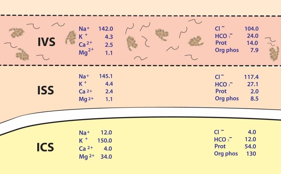
Within each of these spaces are various concentrations of electrolytes (e.g. Na+, K+, Cl, HCO3), along with proteins, glucose, and other substances (Figure 4). Water flows freely between all of these compartments depending on concentration and pressure gradients. Particles will move between these spaces depending on their size, charge, concentration gradient, and pressure gradient.

image13

Figure 4 Normal distribution of particles in the intravascular (IVS), interstitial (ISS), and intracellular (ICS) spaces. Electrolytes (Na+, K+, Cl-, etc.) measured in meq/L.

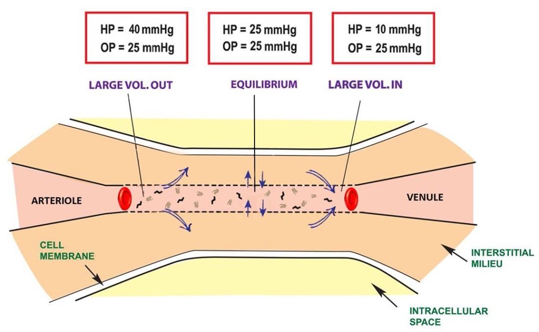
Within the extracellular space, the primary cation is Na+ and the primary anions are Cl and HCO3. These particles flow quite freely between the intravascular and interstitial spaces based on concentration and pressure gradients (Figure 5: as discussed in chapter 1). The cell membrane is a lipid, hydrophobic, barrier. Charged particles cannot cross this barrier without going through specialized pores or channels in the cell membrane. In this way, the intracellular environment can be, and is, vastly different than the extracellular milieu. The primary intracellular cation is K+, and the primary anions are organic phosphates and proteins. Much of the body’s energy (basal metabolism) is spent on maintaining this Na+/K+ gradient by way of providing ATP’s to the sodium/potassium pumps in the cell membrane.

image14

Figure 5 Two opposing forces affect the flow of fluid across the capillary membrane: hydrostatic pressure and oncotic pressure. Hydrostatic pressure drops as the blood travels from the arteriolar end of the capillary to the venule end. Oncotic pressure stays relatively the same. At the midportion of the capillary, under normal conditions, there is equilibrium of pressures such that the only forces that affect particle movement is concentration gradients. At the arteriolar end of the capillary hydrostatic pressure is greater than oncotic pressure and fluid travels from the IVS to the ISS. At the venule end, oncotic pressure from proteins in the plasma is greater than hydrostatic pressure and thus fluid is drawn back into the intravascular space.

Acid /Base Physiology:

Whether a solution is acidic or basic depends on the activity (or concentration) of hydrogen ions (H+) in that solution. The pH of a solution is a measure of its acidity. The human body, through normal metabolism, creates two substances that lead to elevated H+ concentrations: carbon dioxide (CO2), by the metabolism of carbohydrates and fats, and H+ directly, as a result of protein and fatty acid metabolism. The carbonic acid equation explains this relationship between H+ and CO2. H2CO3 is a fairly unstable intermediate molecule, and an equilibrium is simply reached in the body between H+ and CO2 by this equation. HCO3 is a weak base, is far more abundant than H+, and acts as a major buffering system for the body. The abundance of bicarb explains why the normal pH is slightly alkalotic.

The normal pH of plasma in the human body is 7.40 (with a range of 7.35 – 7.45). The body maintains its pH within a very narrow range because of a complex buffering system that it has. As stated, the major buffer in the body is bicarbonate. “Bicarb” too is in equilibrium with carbon dioxide (CO2) and is explained by way of the carbonic acid equation:

image03

Notice that conditions that move this equation to the right (e.g. hyperventilation, which releases more CO2 from the blood) will result in a decrease in H+ concentration, and an increase in pH. Conditions that move the equation to the left (e.g. hypoventilation, which retains CO2 in the blood) results in an increase in H+ concentration, and a decrease in pH. This relationship is mathematically explained by the Henderson-Hasselbalch equation (pK’a is a constant that varies with temperature, and for human physiology is 6.1). We won’t derive this equation further, but note that pH has an inverse proportion to CO2 and a proportionate relationship to HCO3.

pH = pK’a + log10 ([HCO3] / 0.03 * pCO2)

There are 2 major systems at work to control pH in the body: the respiratory system and the renal system. The respiratory system influences the partial pressure of carbon dioxide (pCO2), and the renal system controls the concentrations of H+ and HCO3 in the serum. Disorders in acid/base, then, can result in acidosis or alkalosis. If the disorder is from a malfunction in the management of pCO2, it is referred to as a respiratory disorder, and if the disorder is a result of changes in concentration of H+ or HCO3, it is referred to as a metabolic disorder.

Laboratory Studies:

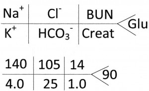
As you may have guessed by now, in order to evaluate a patient with an acid/base or fluid and electrolyte disorder, the physician needs to obtain some lab studies. The following are some basic lab tests that provide a wealth of information as to the status of a patient’s metabolic and respiratory condition. Many abbreviations are used in medicine to expedite charting and the dissemination of information. With regard to electrolytes, a typical “panel” of labs include concentrations of sodium (Na+), potassium (K+), chloride (Cl), and bicarb (HCO3). In addition, this “chemistry panel” typically includes blood urea nitrogen (BUN), creatinine (a breakdown product of muscle), and glucose as well. Common shorthand for displaying this panel is written as in the table below. Notice that the units are omitted because they are of a convention that is utilized widely in labs across the country and the world. For completeness, the approximate average normal values are listed first with their proper units, and then an example is displayed in the table below.

Na+       140 meq/L

K+            4.0 meq/L

Cl-            105 meq/L

HCO3     25 meq/L

BUN        14 mg/dL

Creat        1.0 mg/dL

Glucose     90 mg/dL

image00

Figure 6 Shorthand for a basic chemistry panel

Another very useful test that is performed in order to assess acid/base status in the body is an arterial blood gas (ABG). This sample is drawn from an artery, commonly the radial artery at the level of the wrist, and the partial pressures of oxygen and carbon dioxide (in mmHg), along with the pH of the blood, are measured using a special instrument. A bicarb concentration is also generated, but this is a calculated value based on the Henderson-Hasselbalch equation. The standard format for an ABG report is:

pH/pCO2/pO2/HCO3

A typical normal ABG would be written as:

7.40/40/95/25

The normal values of an ABG with conventional units are:

pH 7.35 – 7.45

pCO2 38 – 42 mmHg

pO2 95 – 100 mmHg

HCO3 24 – 28 mmol/dl

Introduction & Featured Patient Scenario

The Renal System

“Life is like riding a bicycle. To keep your balance you must keep moving.”*

Albert Einstein

Introduction:

Welcome to the toughest clinical topic in the Exploring Medicine course. This is a fun and challenging subject, in that it will bring relevance to some of the biology and chemistry that you have learned in high school and college. If you have been around hospitals much or even watched medical shows on TV, you have probably heard terms like D5W, normal saline, bicarb, and arterial blood gas (“ABG”), among many others. These terms are all references to fluid and electrolyte, and/or acid/base balance. As always, we will start with a presentation of a challenging clinical case; we will then pursue a limited discussion of relevant anatomy, physiology, and pathophysiology. We will conclude this chapter with a return to our case scenario and discuss the diagnosis and treatment of this patient’s disorder and other related disorders.

Patient Scenario

A 42 year old male, with a long history of alcohol abuse, is down on his luck. He has lost his job and now has no money. Without money, he can’t buy booze. If he could obtain any booze a well meaning friend, with whom he lives, would take it away. While sitting in his apartment, he is preoccupied by the fact that he needs alcohol. He can feel the uncomfortable symptoms of withdrawal coming on: shakes, sweats, tremors, abdominal pain, vomiting, muscle cramping, heart racing. He remembers that he has some alcohol in a camp stove that he used a year ago when a friend invited him to go camping. He digs through a cardboard box and finds a can of camping fuel. Being desperate to feed his addiction, he begins to consume this fuel. An hour later, the patient’s roommate arrives home to the apartment and finds the patient comatose. The friend dials 911 and the patient is brought to the emergency room where you are the doctor on duty. What do you do (take a moment to jot down some ideas)?

Natural Science Module Renal

The Renal System

“Life is like riding a bicycle. To keep your balance you must keep moving.”*

Albert Einstein

Introduction:

Welcome to the toughest clinical topic in the Exploring Medicine course. This is a fun and challenging subject, in that it will bring relevance to some of the biology and chemistry that you have learned in high school and college. If you have been around hospitals much or even watched medical shows on TV, you have probably heard terms like D5W, normal saline, bicarb, and arterial blood gas (“ABG”), among many others. These terms are all references to fluid and electrolyte, and/or acid/base balance. As always, we will start with a presentation of a challenging clinical case; we will then pursue a limited discussion of relevant anatomy, physiology, and pathophysiology. We will conclude this chapter with a return to our case scenario and discuss the diagnosis and treatment of this patient’s disorder and other related disorders.