Exploring Medicine – A Foreword

I remember clearly the first meeting I had with Steve Jameson in summer 2005. I had just returned from a regional meeting of the National Association of Advisors in the Health Professions (NAAHP) where a number of sessions had focused on the importance of having health professions advisors build partnerships with health care practitioners in the community. So, when approached by Steve I was more than glad to meet to discuss what I thought was merely an opportunity to enhance our students’ access to shadowing opportunities.

Although that original meeting has led to much greater student access to clinical experiences, then I had no idea that it was the launching pad for an innovative educational experiment in the health sciences that has deeply contributed to the academic, professional, and personal development of hundreds of students – Exploring Medicine. In our first conversations in 2005, Steve proposed to teach a course at the College of Saint Benedict/Saint John’s University (CSB/SJU) that would show students how to critically think like a doctor and how to apply the material they were learning in basic science courses to the process of clinical diagnosis.

We eagerly supported Steve’s idea and in spring 2006 Exploring Medicine was taught for the first time. Since then it has been taught every spring at CSB/SJU and more recently in the fall semester at the University of St. Thomas. Exploring Medicine is truly a gem since it allows students to critically engage in a focused and structured learning experience from a clinical standpoint. Through Steve’s interactive lectures students develop analytical skills that allow them to experience the thinking process of a clinician making a diagnosis; through the panels of healthcare professionals and guest speakers students gain an appreciation for the diversity of the healthcare field and the necessity for a team-approach in care; and last but not least, through the structured shadowing experiences provided by the class students can see how principles covered in the course are directly applied in the process of diagnosis and patient care. Indeed so many students continue to pursue the relationships they have established with clinicians in Exploring Medicine that at CSB/SJU, with Steve Jameson’s help, we established a year-long internship program at the St. Cloud Hospital Emergency Department entitled the Student Health Assistant Program. Engaging community physicians to teach at local universities pays dividends, and Exploring Medicine is the ideal platform to establish those relationships.

The uniqueness of Exploring Medicine is that it is not static – Steve Jameson is continuously editing and tailoring his presentations, creating new experiences for students, and providing novel leaning tools and settings. Since 2006 Exploring Medicine also came to include this book – it is used for the class, but many Exploring Medicine alums will also vouch for its continued value as a refresher and review tool in their further studies. More recently Steve has developed online resources that allow Exploring Medicine to be delivered in its unique and creative fashion to health professions students in any college campus throughout the country; tools that were recognized by the AAMC with its 2013 iCollaborative award in biology.

In short, Exploring Medicine is much more than a course, a book, or a set of online tools. Exploring Medicine is a unique experience that allows health professions students to directly bridge their academic background to a structured clinical setting, and to begin to experience the intellectual world as seen through the eyes of a physician. In the process of helping Steve implement his vision at CSB/SJU, I have seen hundreds of students who are now working as physicians, physician assistants, physical therapists, among others engage in their first meaningful clinical discovery in that setting. Exploring Medicine is indeed the bridge from the world of the humanities, social and natural sciences of our college campuses to the experiential setting of clinical medicine and practice.

Manuel Campos, Ph. D.

Professor of Biology, Preprofessional Health Advisor College of Saint Benedict|Saint John’s University

Preface

Choosing a career is one of the most important decisions you will make in your life. While a career in health care, and in medicine in particular, can be incredibly exciting and rewarding, the journey to that end can be an enormous physical, emotional, and financial drain. The decision to go into this field must be an informed one.

Many are enamored with the medical field and with “being a doctor” long before they know much at all about the practice of medicine. Some are told, “You’re smart; you should be a doctor.” Others simply like what they see on TV. The Exploring Medicine series of modules will primarily focus on what it is like to be a physician, but the information is relevant to all that are seeking a career as a health care professional. To make an informed decision regarding a career in medicine, you should first explore the medical field by seeing what physicians do and learn to think like a physician.

This module, and others in this series, will allow you to do just that. Starting with the very first topic, you will plunge into the world of clinical practice. There you will find patients with a variety of medical problems (based on actual cases), many with life-threatening and life changing emergencies that you will need to work through and solve in order to save your patient’s life and make them well. By the time you have finished this module you will have learned to think and problem-solve like a doctor, and you will understand the process of diagnosing and treating disease.

The medical model is all about diagnosis and treatment of disease, and in order to understand that you have to understand pathophysiology. Pathophysiology is the study of disease. The pathophysiologic process of a disease is the series of events that take place and conditions that develop that result in a particular disease. In order to treat disease, the physician must understand something about the pathophysiologic process that led to the development of that disease. By going through the systems based modules you will gain insight into the thought process physicians use to diagnose and treat disease, and how they help patients to become well. Each system based module begins with a general overview followed by a discussion of the pathophysiology of a particular disorder: e.g. the Respiratory module reviews anatomy and physiology of the Respiratory System, then goes into a discussion regarding the cause of asthma and its treatment. Other modules, like Social Determinants of Health, focus on wellness as it relates to society, social factors, and public policy. Exploring Medicine modules were made for the students that have taken biology, chemistry, sociology, psychology, and other science courses, and wondered, “Why do I have to learn this stuff?” These modules apply the student’s science knowledge to the real world by making correlates to clinical medicine.

Books have been written on what will be small elements of the Exploring Medicine modules, so we obviously cannot comprehensively cover every topic. The authors have distilled a massive volume of material down to several manageable lessons. Despite this editing, the amount of subject matter may still seem intimidating at first, but that is not the intent of the authors. Do not get stressed or anxious. The focus of these modules will be on general principles and concepts. In order to transition from simple anatomy and physiology to treatment of disease, for example, we must streamline our approach in order for the reader to get some appreciation of what it is like to be a physician. The following module will take you, the medical enthusiast, through a very narrow slice of medical training: from medical school, to residency training, to clinical practice. Armed with your newly found knowledge, you will be able to make clinical decisions based on true-to- life patient scenarios and help your patients become well or stay well. If you find this process compelling, as the authors do, then you may seriously want to pursue the exciting field of medicine.

Introduction to the Chapters – Life and Death, Homeostasis and Equilibrium

Death is not the enemy but occasionally needs help with timing. – Peter Safar, M.D. The “Father” of CPR

What is life? That may seem like a metaphysical question, but it’s actually not. It is a very real question for those that provide medical care, but the answer can be surprisingly complicated. As a physician, physician assistant, nurse, or nurse practitioner, you will likely have many occasions where you will be dealing with patients as they look to cross that line from life to death. In the following chapters we will discuss a variety of pathophysiologic processes that will alter the normal function of the human body.

Before we delve into specific disease processes, though, let’s take a look at a more fundamental principle of human physiology and how a disruption of normal processes can lead to chaos, i.e. entropy, on a microscopic (actually molecular) level and to death on a macroscopic level.

The human body is a complex network of systems that operate in unity to keep us alive and active. In order to maintain normal function the body seeks homeostasis and defies equilibrium. Cellular reactions take place under very specific conditions: a narrow range of pH, the correct concentrations of particular ions, proper amount of substrates, etc. Homeostasis is the set of conditions that the body establishes and maintains in order to function properly. It takes energy to maintain this homeostasis. This energy has to be consumed in the form of chemical bonds contained in the food we eat, converted to a usable form, transported throughout the body to all of its cells, and metabolized into a form of energy the cells can use: ATP. The wastes of these reactions then have to be removed. It is a complex network of integrated systems that accomplishes all of these tasks.

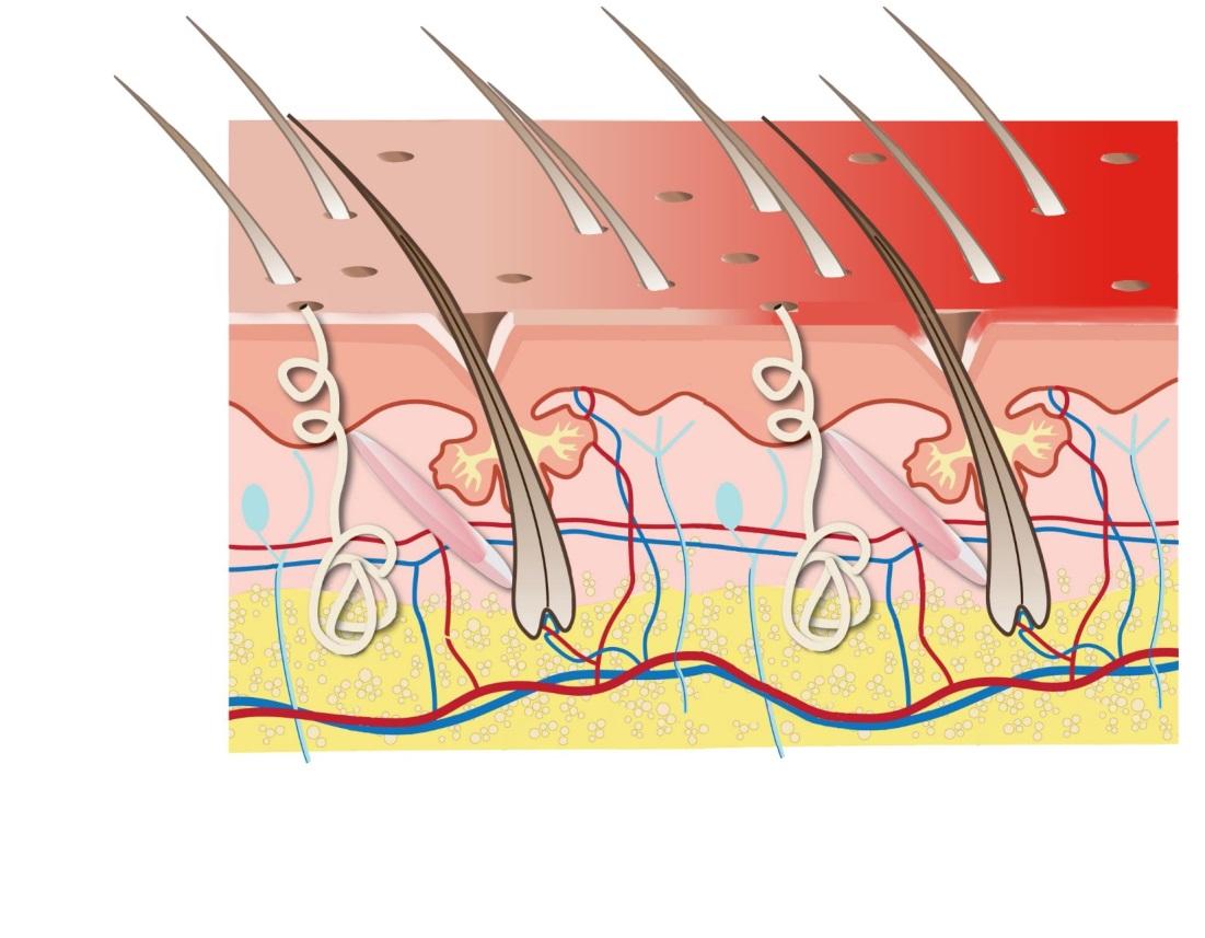
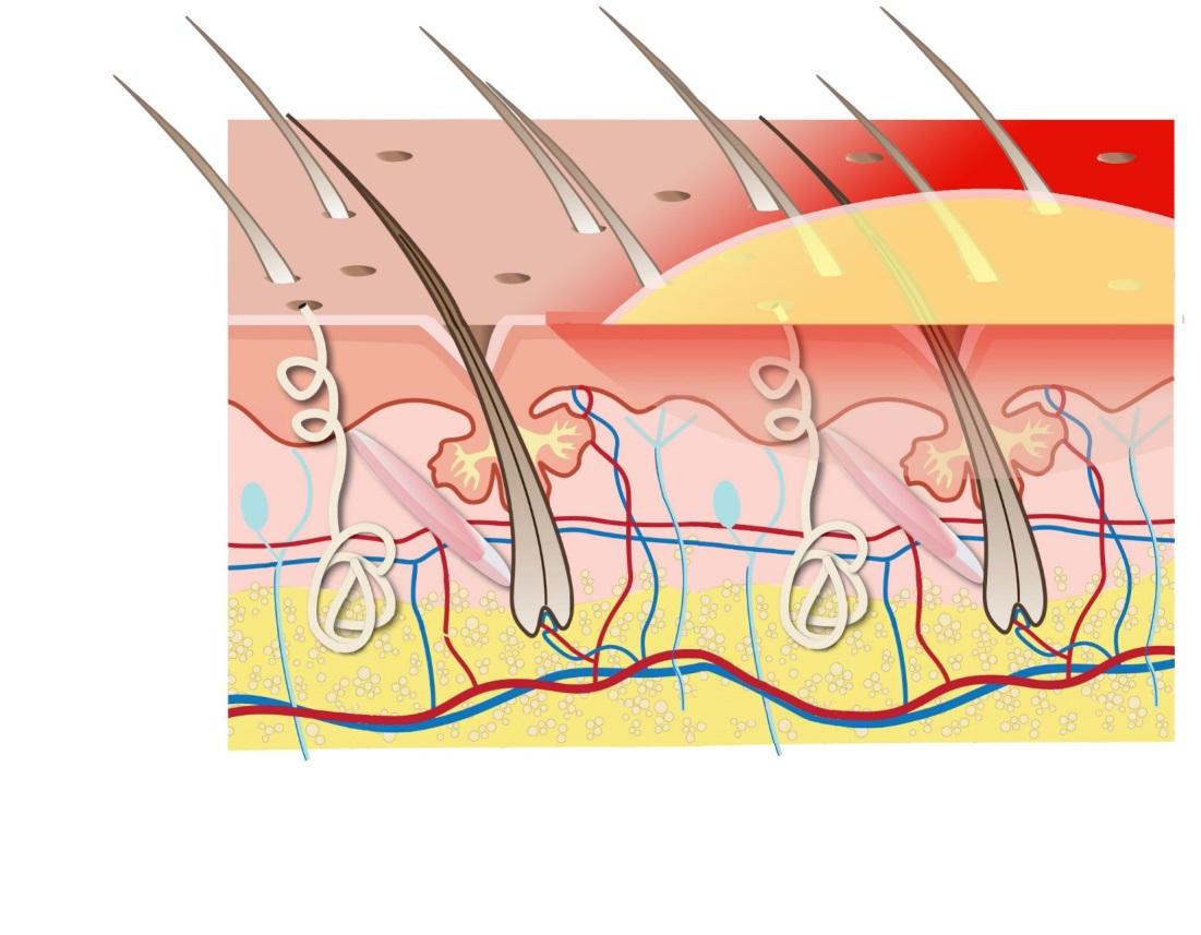
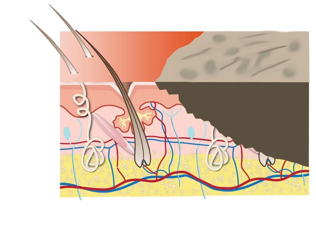
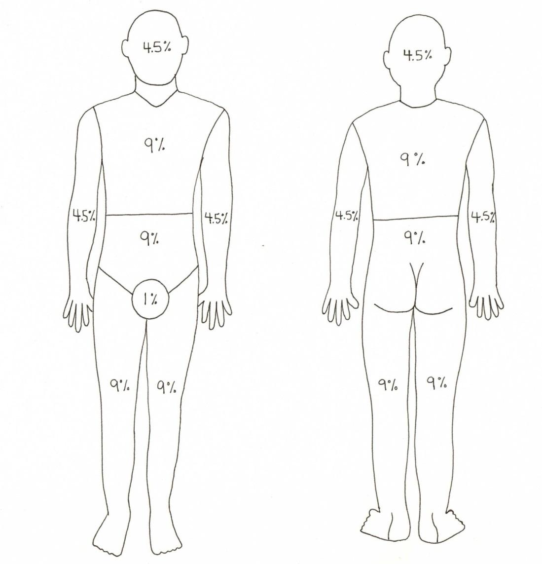
In Exploring Medicine, we will look at four of the body’s integrally related physiologic systems (Cardiovascular, Respiratory, Renal and Integumentary) as examples of how the body maintains homeostasis. We also explore what can happen when these systems fail, and what we, as medical care providers, do to restore normal function and reverse the process of dying. All of the chapters have questions to answer along the way and challenging scenarios to solve at the end of each chapter. Answers to these questions and further elaboration of concepts are in the appendices near the end of the book. In the chapter on Evidence Based Medicine, we will discover how the medical literature helps to guide a physician’s clinical practice. As you strive to learn more about the field of medicine, remember the words of Hippocrates (the final line of the Hippocratic Oath), “May I always act so as to preserve the finest traditions of my calling and may I long experience the joy of healing those who seek my help.”

Integumentary System Unit Quiz

This is an open book test.  You can click here to open the lesson in a new tab/window to review the lesson as you take the quiz.  If you close the quiz you will have to start all over with a different set of questions. You need to achieve a score of 90% or greater in order to obtain the certificate of completion.

Please ensure you have enough time to complete the entire quiz at one time.

Other Clinical Integumentary Scenarios

The following scenarios are designed to walk the future physician through a variety of real life situations that highlight disorders of the integumentary system. Read the introduction closely and work through the scenario to ultimately diagnose and treat this patient’s condition.

Clinical application: Our burn patient

In the emergency department, oxygen is being given by face mask and an intravenous line has been inserted. Our patient has pale and charred skin on the scalp, face, torso, arms and hands, and upper legs. She is spared any burns below the knees and there are small areas of sparing on the torso. After a brief examination, you order some potent intravenous pain medication. At the same time, you prepare for ambulance transportation to a specialized burn unit, which in this case is eighty miles away.

There are many issues that need to be addressed and treated before transport occurs. She first needs her airway and breathing protected and she needs pain control. This necessitates endotracheal intubation and assisted ventilation. Large doses of narcotic pain medications are given. Recall, as well, that one of the major consequences of major burns is fluid loss. In our Renal chapter we learned that we use an isotonic solution such as “normal saline” to expand the intravascular space and that is what we will use for fluid replacement here. Our colleagues at in have developed a formula for fluid resuscitation in burn victims and it is as follows: take the percent body surface area that has sustained second and third degree burns, multiply this number by the patient’s weight in kilograms, and then multiply this product by 4 ml. This is how much fluid that the patient needs to receive in the first 24 hours. Her burn is 80% BSA and her weight is 50 kg, therefore she needs to receive 16 liters of isotonic fluid in addition to her maintenance fluid in the first 24 hours.

Parkland Formula for fluid resuscitation in burns:

24 hour fluid requirement = weight (kg) X %BSA of burn X 4 ml/kg

24 hour fluid requirement = 50 kg (80) (4ml/kg)

24 hour fluid requirement = 18.1L

Understandably, the girl’s family is stunned by the event, and wants to know why she can’t stay at their local hospital. They also want to know if she will be okay and if she will have any long term problems. You provide an accurate yet compassionate assessment of the situation.

Reviewing the pathophysiology and treatment of burns above, it is clear that this patient has a major life-threatening burn. In a young healthy patient, survival is possible but certainly not guaranteed. You discuss with the family the need for highly specialized care of the burn itself, including dressing and skin grafting, along with the intensive care to deal with the fluid losses and severe infections that commonly accompany these injuries. In addition, you discuss the need to place an endotracheal tube prior to transfer in order to protect the airway and permit more aggressive sedation and pain control. And yes, there will be scarring, but skin grafts and the use of cultured skin, as well as reconstructive procedures will go a long way towards restoring an acceptable cosmetic outcome. This event will change her life in many ways, but she will still be the same wonderful person she was before.

Pathophysiology & Treatment of Burns

A burn is an injury to the skin that occurs as a result of excessive heat (thermal burn) or as a result of exposure to a chemical (typically a strong acid or base). Burns are categorized by the “degree” in which the skin is damaged, and by the percentage of body surface area involved. We will focus on thermal burns as it relates to our patient scenario. In addition, the treatment of thermal burns is straightforward. Thermal burns are generally broken into 3 categories: first degree, second degree, and third degree.

A first degree burn is a superficial burn, involving only the most superficial layer of skin: the epidermis. Mild sunburn (no blisters) is a good example (Figure 6). The skin is red, blisters are absent, and important structures are spared. While these injuries can be painful, healing without long term consequences is the expected result, even in a burn involving virtually 100% of the body surface area.

image17

Figure 6 Cutaway view of the skin: first degree burn. The injury is limited to the superficial layers of epidermis and dermis.

Second degree burns (Figure 7) involve deeper layers of the skin. Superficial layers separate and form fluid-filled blisters. In a large burn, more than 15 – 20 % of body surface area, enough body fluid can be diverted into these blisters to cause hypovolemic shock. When the blisters rupture, the area becomes susceptible to infection. Because the deeper layers containing the nerve endings are spared (not damaged), second degree burns are very painful. With partial sparing of the growth layer, most of these burns will heal with new skin rather than scarring.

image18

Figure 7 Cutaway view of the skin: second degree burn. This injury involves deeper layers of the dermis. The deepest layers and important structures – nerves, blood vessels, sweat and sebaceous glands and hair follicles — remain intact. Remnants of the germinal layer remain intact, conferring potential for healing by growth of new skin.

Third degree burns (Figure 8) destroy the entire thickness of the skin. Because nerve endings are destroyed, the burn is painless, but the consequences are more severe. The germinal layer of the epidermis is destroyed in this full thickness burn; therefore the body is incapable of growing new skin in the affected area.

image19

Figure 8 Cutaway view of the skin: third degree burn. This is a full-thickness injury. Nerves are destroyed, so the wound is painless. The growth layers of the dermis are also lost, so healing will occur by scarring rather than by growth of new skin.

Fluid loss and infection can be life-threatening and victims of a major burn may spend weeks or months in the hospital. Among patients sustaining a severe burn to 80- 90% of the total body surface, only half will survive.

Because the burn heals by scarring rather than re-growth of skin, those who do survive are deprived of the protective, heat-regulating, elastic tissue that constitutes an important interface between us and our environment.

That brings us to the next issue regarding burns and that is the amount of skin involved. Besides depth, the other crucial factor regarding burns is how much surface area of skin has at least second or third degree burn involvement: this is referred to as the percent of total body surface area (BSA). Estimating this can be difficult but a good method is to use the “rule of 9’s” (Figure 9). For smaller burns we use the patient’s palm (not including the fingers or wrist) to estimate how many “palm sizes” a particular burn is. Each palm size is nearly 1% of BSA.

image14

Figure 9 The “Rule of 9’s”. The numbers are added to determine the percent of total body surface area that is burned.

Treatment of Burns:

Simple burn care consists of cleaning and dressing the wound. The biggest concern for the patient, however, is pain control. There is nothing like the pain of a severe second degree burn, and there is nothing like the instant relief one achieves by simply running the affected area under cold water or placing the burned area in a cool water bath.

image15

Figure 10 This is a deep second degree burn, the result of a lighter that exploded in this poor gentleman’s hand. Opiates are the mainstay of medications for pain control, after cool water. Anti-inflammatories (e.g. ibuprofen or naproxen) are also used as an adjunct for pain control. After reasonable pain control is achieved, dressing the wound with antibiotic ointment applied under a gauze dressing would be appropriate. The patient needs a one day follow-up to confirm the depth of the wound: which often looks more superficial on the first day.

When dealing with serious burns involving large surface areas of skin, it should come as no surprise that airway and breathing issues are high priority, as they are in all medical emergencies. Swelling in the throat, face, and neck can compromise the airway. Smoke inhalation can severely compromise the ability of the lungs to oxygenate blood, and loss of elasticity of skin over a chest burn can prevent normal chest expansion with inspiration. For these reasons, supplemental oxygen is commonly necessary, as is endotracheal intubation and assisted ventilation.

Hypovolemia due to fluid loss through the burn is the next major priority. Patients with large areas of third degree burns may require many liters of intravenous fluid every day for the first few days in order to replace these fluid losses.

While the region of the third degree burn itself isn’t painful, it typically coexists with areas of first and second-degree injury: both of which are very painful. Burn patients require large doses of strong pain medication.

Care of the burn itself is, surprisingly, not an early priority in major burns, but is crucially important once the ABC’s have been addressed. While less serious burn wounds can be treated with antibacterial ointments and bandages, larger ones require cleaning in whirl pools, coverage with more specialized dressings, and grafts from the patient’s own skin and/or cultured skin (grown in the laboratory).

Clinical Application: Our acne patient

Recall that our patient, a 15 year old girl, has moderately severe acne and is suffering emotionally because of this. Obviously we need to help this girl by treating her acne, but let’s take a moment to address her psychosocial issues (the girl’s ability to cope with her disease). Why did this girl go into such a serious downward emotional and social spiral from such a benign disorder? After all, acne is just pimples and it should be a simple problem to assess and treat – shouldn’t it? The answer is no, acne is not always a simple problem and it can leave both physical and emotional scars. Acne commonly occurs on highly visible areas such as the face, upper chest, and upper back, and can greatly affect self-esteem as it tends to disfigure these areas.

Regarding the patient’s acne, she has already tried a variety of soaps and even cleansers containing benzoyl peroxide. She is not seeing much success with this “first line” treatment. In an effort to minimize potential adverse effects, yet give her the best chance for success, what treatment plan would you recommend at this point?

Recall that estrogens (a major component of OCPs’) tend to suppress sebum production and frequently lead to an improvement in acne. Oral contraceptives could potentially regulate her menstrual cycle as well as prevent an unwanted pregnancy. Discussing the risks and benefits of this therapy would be prudent.

In addition to OCP’s, you will want her to continue using the antibacterial soap and benzoyl peroxide that she is already using. Another therapy to consider is antibiotics. You could start with topical antibiotics but most clinicians would probably go right to oral antibiotics with this particular patient.

“Surgical” treatment such as comedone extraction could be considered at this time but injections with anti-inflammatories and laser and dermabrasion would not be indicated this early in the treatment of this disease. Treatments like these are expensive and medical insurance plans offer minimal coverage for procedures considered “cosmetic” — performed primarily for reasons of appearance.

You discuss the treatment plan with the patient and her mother. You assure the girl that you are taking her acne problem very seriously and that you are going to see her back in just a few weeks to be certain she is doing okay. She knows that even if her acne is not controlled at that point that you have other therapies to offer. Both mother and patient leave feeling pleased.

Just then, a call comes in from the emergency department. They have just admitted a 12 year old girl with burns estimated to cover 80% of her body. She had been at a friend’s for a sleepover. They built a fire in order to roast marshmallows. New wood thrown on the ash wouldn’t light again this morning, so the patient grabbed a small can of gasoline and poured it on the fire. Immediately the flames rushed up the stream of gas, exploded gas into the air and on the patient, and ignited her hair and clothes. She ran to the house, and the father of the girl’s friend, hearing the commotion, ran to the patio to see what the matter was. He grabbed the young girl, now engulfed in flames, and rolled her in the grass to extinguish the blaze.

In the emergency department you find the girl to be quite stoic but clearly in pain. She is breathing on her own and is not coughing. She has pale and charred skin on the scalp, face, torso, arms and hands, and upper legs. She is spared any burns below the knees and there are small areas of sparing on the torso. What do you do?

Pathophysiology & Treatment of Acne

There are actually a number of types and classifications of acne and related conditions, but for this discussion we will concern ourselves with acne vulgaris (vulgaris = common). As the name implies, this condition is encountered frequently but it is anything but simple. Among the causes of acne are hormonal influences, genetic predisposition, contact with skin (e.g. touching face with dirty hands or use of chin straps on helmets), and use of certain medications.

Interestingly, particular foods like potato chips and chocolate, long thought to be a cause of acne, have not widely been associated with inducing this skin disorder. Among its consequences are the development of more serious skin infections, scarring, embarrassment, social isolation, and other psychological trauma. Acne treatment has become a multi-billion dollar industry.

Typically, acne starts when the sebaceous glands are induced to produce more oil. This commonly happens during puberty, when levels of sex hormones, especially testosterone, rise. (Testosterone is usually considered a male hormone, but females have small amounts of this as well). Under the influence of these hormones, sebaceous glands enlarge, and the increased sebum mixes with sloughed lining of the follicle and gets clogged at the neck of the hair follicle. This plug is the typical early mark of acne, the comedone. When the pore remains closed with the sebaceous plug, this is referred to as a closed comedone, commonly called a “white head.” When the orifice of the plugged pore enlarges as cellular debris accumulates and darkens, an open comedone or “blackhead” develops. As bacteria propagate behind this obstruction and in the rich environment of sebum, inflammation occurs and a papule develops. Further inflammation and the production of pus, from the body’s immune response to the infection, leads to the formation of pustules (Figures 3 and 4a-e).

image07

Figure 3 Progression of an acne lesion: an overview (Used with permission of Galderma Laboratories, Fort Worth, TX)

image08

Figure 4a Normally functioning hair follicle and sebaceous gland (Used with permission of Galderma Laboratories, Fort Worth, TX)

image09

Figure 4b Development of the microcomedo: the neck of the hair follicle becomes plugged by skin debris (from excess keratinization – sloughing of the outer layer of the lining of the follicle) mixed with sebum (which is produced in excess). (Used with permission of Galderma Laboratories, Fort Worth, TX)

image01

Figure 4c Progression of a closed comedone (“whitehead”) to an open comedone (“blackhead”). This occurs as the orifice of the pore enlarges and accumulates additional debris. (Used with permission of Galderma Laboratories, Fort Worth, TX)

image00

Figure 4d Progression from comedone to papule, resulting from infection and inflammation (Used with permission of Galderma Laboratories, Fort Worth, TX)

image03

Figure 4e Further progression from papule to pustule, with pus visible above the skin surface. (Used with permission of Galderma Laboratories, Fort Worth, TX)

image02

Figure 4f Papular and early pustular acne

When a hair follicle gets plugged, sebum accumulates behind the obstruction because the sebaceous gland keeps producing this oily substance. One particular bacterial species, Propionibacterium acnes (P. acnes), flourishes in this environment. The human body recognizes the rapid proliferation of bacteria in this sebum filled, occluded follicle and sends white blood cells (WBC’s) to fight this infection. These WBC’s release a variety of inflammatory mediators and this results in redness and swelling. As more WBC’s are drawn into the follicle they, along with the bacteria they kill and other cellular debris, accumulate as pus. As more pus develops within the follicle, the surrounding area becomes more reddened and swollen and even warm and tender. The resulting pustule is commonly referred to as a pimple. The longer and more intense this inflammatory reaction is, the more the lesions progress. Though small at first, these pustules can grow to become large cysts, and these cysts can coalesce and form fistulous tracts between them and result in severe deformity and scarring of the involved areas of skin (Figure 5a-g).

image04

Figure 5a Hair follicle and sebaceous gland: early accumulation of sebum (yellow). The follicle is intact. The small bubble insert shows an enhanced microscopic view of the follicle lining.

image10

Figure 5b As pus (white blood cells and bacteria) accumulates within the follicle, pressure rises and the wall of the follicle begins to bulge.

image11

Figure 5c When the pressure within the follicle reaches a critical point, rupture of the follicular wall occurs and sebum, white blood cells, and bacteria are released into the subcutaneous tissue. This attracts scavenger cells known as macrophages (green) to help clean up this infection.

image12

Figure 5d Common acne cyst

Unfortunately, acne commonly affects some of the most visible areas on the body. Scarring is the worst (physical) long term consequence of acne (Fig. 5f). In the past it was often unavoidable, but now there are a variety of treatments that may control the acne before it reaches the scarring stages, as well as procedures that can restore scarred and irregular skin to a more normal appearance.

image13

Fig 5e Another acne cyst in background of more serious cystic acne

image16

Figure 5f Scarring as a result of severe cystic acne

Treatment of Acne:

As in all science-based medicine, therapeutic interventions are based, whenever possible, on an understanding of pathophysiologic processes. Layered on top of this is knowledge of the specific treatments, both their expected results and potential adverse effects, as determined by clinical research. Finally there is application to the individual patient ideally based on collaboration between patient and practitioner.

In the early stages of acne, simple treatments are often all that is needed. Antibacterial soap is one of the simplest first treatments: it kills the offending P. acnes bacteria and it cleans off excess oil (sebum) from the surface of the skin. Benzoyl peroxide, which can be purchased without a prescription, is another first line treatment. This agent is available in a variety of liquids and gels and mixed in a variety of media allowing it to dry the skin, dissolve comedones, and break down excess sebum on the surface of the skin so it can be easily washed away. It also has some antibacterial activity and is combined with other agents in an effort to make it even more effective. This sort of treatment carries little risk of unwanted effects so it is perfect for an initial treatment. In minor acne this treatment may be all that is necessary.

If these agents are not sufficiently effective, other types of topical treatment can be considered. Derivatives of vitamin A (retinoids) cause shedding of skin cells and reduce the accumulation of excess keratin within the follicle. Topical antibiotics are another second line of defense against acne and they work by decreasing bacterial proliferation. These two agents are commonly used in combination in moderate acne. Both of these agents however, carry some risk of significant skin irritation and allergic reaction.

If the acne reaches the nodular or cystic stage, more aggressive treatment may be considered. Direct care of the lesion by the dermatologist – extracting the sebaceous plugs from comedones or injecting nodules and cysts with anti-inflammatory medications can be helpful in the short term, but they are time-consuming and expensive.

In addition to the topical agents described above, oral medications may be needed as well. Both antibiotics and retinoids come in oral forms, and tend to be more effective when taken in this way. Introducing a drug into the bloodstream, however, generally introduces the potential for more serious side effects as well. The retinoids especially carry a number of known risks, including liver toxicity, sores at the side of the mouth and elevation of undesirable lipids in the bloodstream. The most significant issue with these drugs is that, when taken during pregnancy, they pose serious dangers to a developing fetus by causing birth defects. Since young women in their childbearing years are among the most common types of patients seeking treatment for a cosmetically significant condition, this issue has resulted in strict formal protocols to ensure that these medications are not administered during pregnancy.

And speaking of pregnancy, recall the role of the sex hormones in the production of sebum: testosterone tends to increase the quantity and density of sebum, while estrogens have the opposite effect. Hormonal manipulation, usually by the administration of estrogen as an oral contraceptive, can have a beneficial effect for the patient with acne. For younger women who may desire the contraceptive and menstrual period-regulating effect of birth control pills, this may be a good solution. For women ready to have children, this would not be a good option. Moreover, even such “natural” substances as estrogens can be associated with increased risk of blood clots in the legs, lungs and brain, and possibly some cancers. Hormone therapy is obviously not a viable choice for males since an excessive influence of estrogen would result in gynecomastia (development of breasts) and other undesirable effects.

Finally, what can be done for the patient who, after years of severe acne, ends up with the scarring and uneven skin surface? Fortunately some of these changes can be erased, or at least improved, by processes designed to very carefully eradicate the superficial layers of damaged skin. This is carried out by the use of laser light, chemicals, and abrasives. The expectation is that the healing process will result in a smoother and more uniform appearance to the skin.

Background, Anatomy, Physiology

Skin has many functions that are necessary for survival: it serves as a barrier to infection, it stores and holds fluids and energy, it plays a large role in regulating body temperature, and it contains sensors that allow us to feel the environment around us.

As the largest and most visible organ of the body, skin also plays a prominent role in the perception of physical beauty. Diseases of the skin can lead to embarrassment, loss of self-esteem, and social isolation. Some will deal with debilitating skin disorders better than others. As a healthcare provider, you need to individualize treatments for skin disorders based not only on the patient’s physical condition, but also on the patient’s emotional response to the severity of the condition. Treating acne, for example, seems straightforward but a provider can easily miss the emotional importance of this condition. Conversely, it is not difficult to be lured into aggressively over-treating this disease and lose sight of the fact that there are risks to some therapies that may outweigh the benefits.

Anatomy and Physiology of the skin:

In the average adult, the skin covers an area of almost 2 square meters. Cutis is the Latin word for skin and thus when we refer to structures within the skin we refer to them as “cutaneous” (of the skin) structures. Skin is composed of 2 major layers: the epidermis and the dermis (Figure 1).

image05

Figure 1 Cross-section of the skin: epidermis, dermis, and subcutaneous tissue

The epidermis is the outermost layer of the skin. This is the body’s first defense against external threats such as desiccation, extremes of temperature, ultraviolet radiation, and microorganisms. The epidermis is thin and primarily consists of multiple layers of dead cells that had their origins in the deeper, generative layer. At the base of the epidermis is the basal layer. This single layer of cells is made up of keratinocytes (squamous epithelial cells that form the outer skin and produce keratin) and melanocytes (pigment producing cells that determine skin color). It is the only place in the epidermis that proliferation (cell division for new cell growth) takes place. Beneath the epidermis, the dermis is a more complex layer housing a variety of structures important to the functions of the skin as outlined above (Figure 2). Sweat glands open and close in response to stimuli from nerves and hormones; they help to maintain a constant body temperature. Hair follicles are genetically programmed to produce various types and quantities of hair in different parts of the body and are supplied with the tiny muscles that “make your hair stand on end.” This phenomenon, in other animals, serves to increase the insulating ability of the hair (fur), but in humans is mainly responsible for the phenomenon of “goose pimples.” Closely associated with the hair follicles are the sebaceous glands which secrete the oil that adds to the waterproof properties of our skin. Rather than being evenly distributed over the skin, the sebaceous glands are concentrated in the head, neck and upper torso, which has medical consequences that will be described below.

image06

Figure 2 A small block skin and a depiction of the structures within: nerves, blood vessels, sweat glands, sebaceous glands, hair follicles

Below, or deep to, the dermis is the subcutaneous tissue, composed largely of loose connective tissue and fat. This layer may be considered part of the same functional unit as the dermis and it is physically attached to the skin more than to the underlying tissues. The dermis and subcutaneous tissue serve as the primary insulation of the body.

Having their origins elsewhere, but traveling through the subcutaneous tissue to their destination near the surface of the skin are numerous blood vessels and nerves. The second-smallest branches of the blood vessels, the arterioles, can constrict or dilate to regulate flow of blood into the skin’s capillary bed in order to expose more or less surface to the environment and serve to regulate body temperature. The nerves terminate in specialized endings that serve as transducers for the senses of touch, vibration, and hot and cold.

Pathophysiology of the Integumentary System:

There are a variety of diseases and injuries that can affect our skin. In the paragraphs below, we will highlight a broad range of these conditions, but with a special emphasis on acne and burns.

Because the skin is a major interface between ourselves and our environment, it should not be surprising that the environment can have an impact on the skin. We refer, of course, to trauma of various sorts: lacerations (cuts), abrasions (scrapes), contusions (bruises), and burns would describe most types of injury to the skin. Loss of major areas of skin – for example, a deep burn of more than 40% of the body’s surface – can be life-threatening. Fortunately, in all but the most serious injuries, the skin has a remarkable ability to regenerate and heal, often with little need of medical intervention.

Infections of the skin reflect the interaction of skin with the microbial world outside. While bacteria, viruses, and fungi often live within the epidermis, the term infection connotes an invasive process in which large populations of microbes breach the skin’s defenses. When the skin is invaded by pathogens such as these and the invading organism begins to multiply, it is referred to as cellulitis. This infection can result in destructive processes that progress to loss of a body part or even death. Host defense mechanisms, such as inflammation and fever, also come into play. White blood cells invade the area and form the pus in abscesses and tiny pustules in some skin infections.

Inflammation is a complex phenomenon in which the body’s defense mechanisms produce characteristic changes such as localized warmth, redness, and swelling. While inflammation may be an appropriate reaction to infection and other injuries, it may also occur when it is not wanted. The generic term for inflammation of the skin is dermatitis and there are dozens of kinds skin diseases such as eczema, often associated with allergies and asthma. Poison ivy is an example of a contact dermatitis. In this case, a resin from the plant penetrates the outer layer of skin and the response by the body’s immune system leads to itching, inflammation, and blisters. There is a wide range of skin diseases in which inflammation is a prominent feature. In some cases the inflammation can be so severe as to result in major skin loss and even death.

Several skin problems are the result of a dysfunction of hair follicles. Acne (discussed in detail below) is the most common of these disorders, but there are many other conditions in which the hair follicle gets closed and allows for the development of inflammation and infection within the follicle. These conditions include pilonidal abscess (infected hair follicle that develops into an abscess and is located in the upper central crease between the buttocks), hidradenitis suppurativa (inflammation and abscesses that develop in the folds under the arms and in the groin), and hot tub folliculitis (inflammation that occurs in hair follicles after being in a hot tub and reacting to chemicals and/or infection).

Like any other organ in the body, skin is susceptible to abnormal growths and cancer. Non-cancerous growths are referred to as “benign”, meaning they are biologically programmed not to spread beyond a limited area. Cancerous (malignant) tumors invade local tissues and can spread to other parts of the body. Cancer of the skin is the process in which a germinal cell begins to divide in an unusual manner and at an accelerated rate. This leads to a bump or colored area on the skin. Some cancers grow as a localized lesion (e.g. basal cell carcinoma) until they become very large and others begin to spread (metastasize) throughout the body. One particularly serious form of skin cancer is malignant melanoma, recognized by its dark coloration. Skin cancers, and even benign growths, are often the result of excessive UV exposure from the sun.

In contrast to the cancers, some cutaneous diseases are the result of abnormal growth of normal tissues. Psoriasis is a disorder in which the skin simply grows too fast in some areas and becomes thickened with too much of the outer keratin layer (hyperkeratinization). This is a benign growth but can be very debilitating and disfiguring.

Because the skin is the most visible component of our bodies, many systemic diseases can be diagnosed by their unique prominent dermatologic manifestations. It is common knowledge that varicella (chickenpox) causes a rash consisting of tiny blister-like lesions (vesicles) on a red base. Measles and rubella, now uncommon in the U.S. due to vaccination, cause rashes with classic characteristics: a doctor only needed to see the rash, years ago when it was common, to diagnose this disease. There is a wide assortment of other systemic infections, allergic conditions, and inherited diseases that are accompanied by characteristic skin changes.

Dermatological Definitions:

In all medical specialties, a localized abnormal structure in the body is termed a lesion. Because the field of dermatology is heavily weighted towards the visual, a fairly precise system of terminology has been developed to describe lesions of the skin. For example, a small, non-elevated colored spot is called a macule; when the spot is elevated, it becomes a papule. A tiny superficial fluid-filled lesion is a vesicle, while a much larger one is a bulla. A papule is a small, raised, conical shaped lesion, commonly red and inflamed. When a papule becomes slightly larger and fills with pus, it is called a pustule. A soft, red, raised, blanching (turns skin color when compressed) skin lesion that is pruritic (itchy) and can be quite large with irregular borders is urticaria (hives): typically the result of an allergic reaction. These are not entirely arbitrary categories, as location within the skin structure and specific pathophysiology confer on these lesions their characteristic appearances.

Patient Scenario

As a family physician in a small community, your day started like any other as you went to see the first patient in your office at 8:05 AM. Sitting across from you is a 15-year-old girl for whom you have been caring since birth, but haven’t seen for more than 3 years. The young girl is bent over with her face in her hands, and her mother is tearful with an arm draped around her daughter. The mother states that she just found out last night that her daughter has not been going to classes regularly for almost two months, and has been classified as truant. Previously an honor student, the patient has begun to fail in school and has been ostracized from her group of high-achieving friends. In the interview, you learn that the girl has been developing increasing anxiety and depression over the past couple of years. She has little self-esteem and refers to herself as “ugly.” She raises her head up as she says this and reveals what she says is the source of “all” of her problems. She states that she has been made fun of at school and that nobody likes her. When you closely examine the girl’s face and back, you see numerous pustular and cystic acne lesions, and even several small, pitted scars. This is a moderately severe case of acne. What do you do? Jot down a few ideas of what you think the issues are and what you would do to help this troubled girl.

Natural Science Module Int

The Integumentary System

Years may wrinkle the skin but to give up enthusiasm wrinkles the soul.*

Samuel Ullman

Introduction:

Imagine a high-tech fabric with the following properties: 1) It is waterproof, yet it allows moisture and heat to escape as needed. 2) It is flexible, tough, thick or thin in areas in which those properties are needed. 3) It has friction ridges to prevent slippage, and 4) it has the ability to seal itself when damaged. Does such a fabric really exist?
It does! This miracle fabric is, of course, human skin.