Pathophysiology of Shock

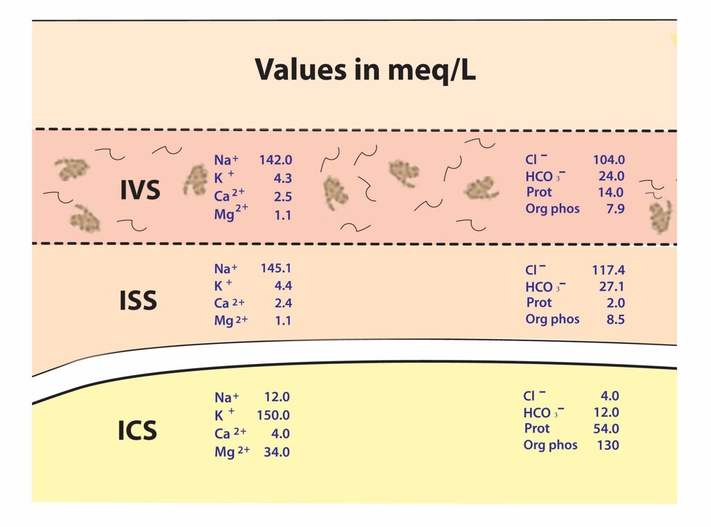
In a normal human physiologic state, there is a homeostasis that exists in cells that includes good stores of energy (e.g. ATP) and energy sources (e.g. glucose), an adequate supply of oxygen, and a pH , fluid, and electrolyte balance that optimizes cellular function. About 20% of the energy expended in the body at rest, that is 20% of the basal metabolic rate (BMR), is used to maintain Na+ and K+ gradients via the Na+/K+ ATPase pump. In fact, the brain and kidneys use 50 – 70% of their BMR on this function.  If any of the conditions above are disrupted, say by lack of blood flow to cells of the body, cellular dysfunction occurs.  If disruption persists for a long enough period of time functions, such as the Na+/K+ ATPase pump on the cell membrane, begin to fail and Na+ and K+ (along with fluid) follow diffusion gradients and begin to equilibrate between the intracellular space and interstitial space.  Homeostasis is lost, enzymes fail to function properly, and cellular death occurs.  The goal, then, is to recognize and treat shock when it first manifests.  This will decrease morbidity and mortality in your patients.

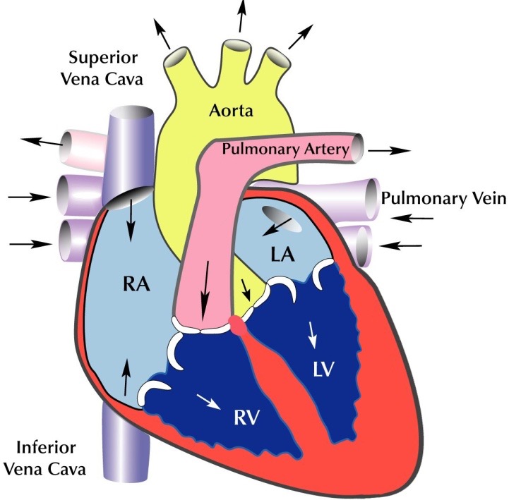
So, what is shock?  Clearly stated, shock is defined as a decrease in circulation of oxygen and/or nutrients to tissues to the point that those tissues malfunction, become damaged, and ultimately, in severe cases, are destroyed.  In order to avoid shock, then, one must have adequate blood flow to the tissues, and that blood has to be carrying adequate oxygen and nutrients.  In resuscitation of any critically ill or injured patient, the physician must address the ABC’s: i.e. Airway, Breathing, and Circulation.  If the patient cannot maintain a clear airway, one must be provided, often by placement of an endotracheal tube. Breathing, if inadequate, must be assisted. If the patient is hypoxic (has low oxygen in the blood), supplemental oxygen is administered as well. The third component of this triad, circulation, is central to the pathophysiology and treatment of shock. 

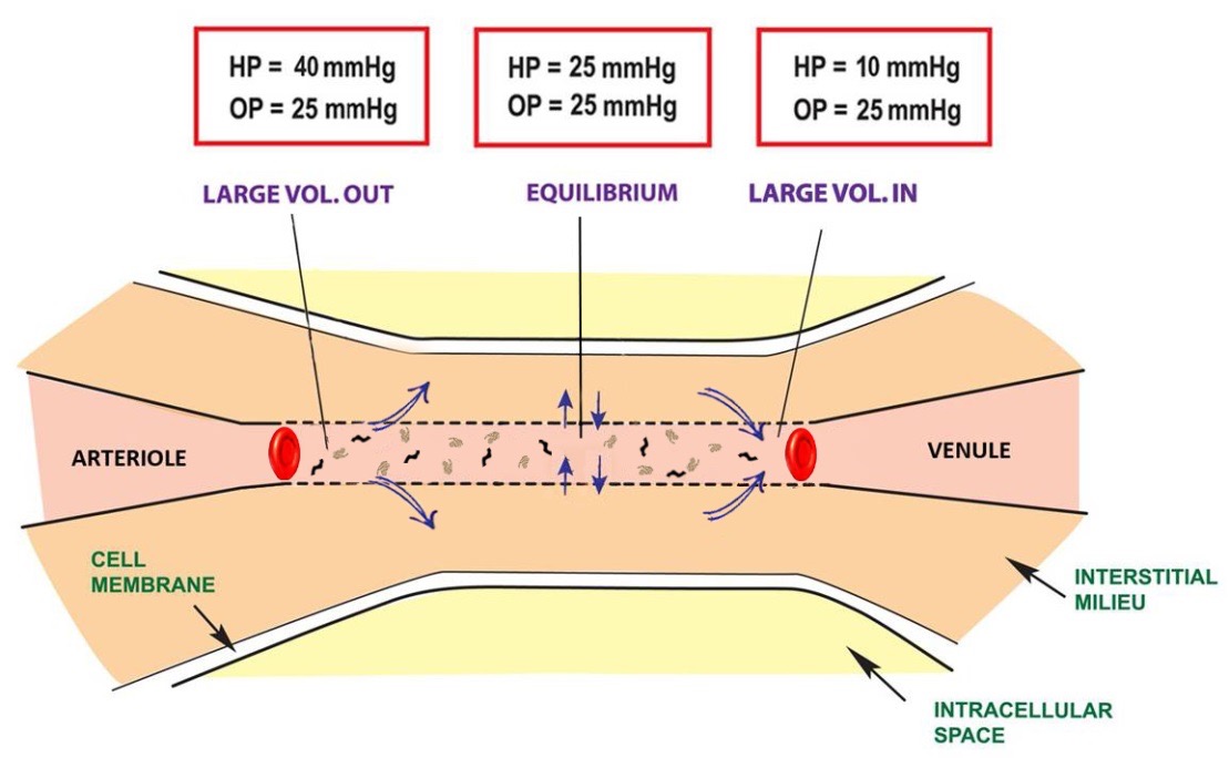
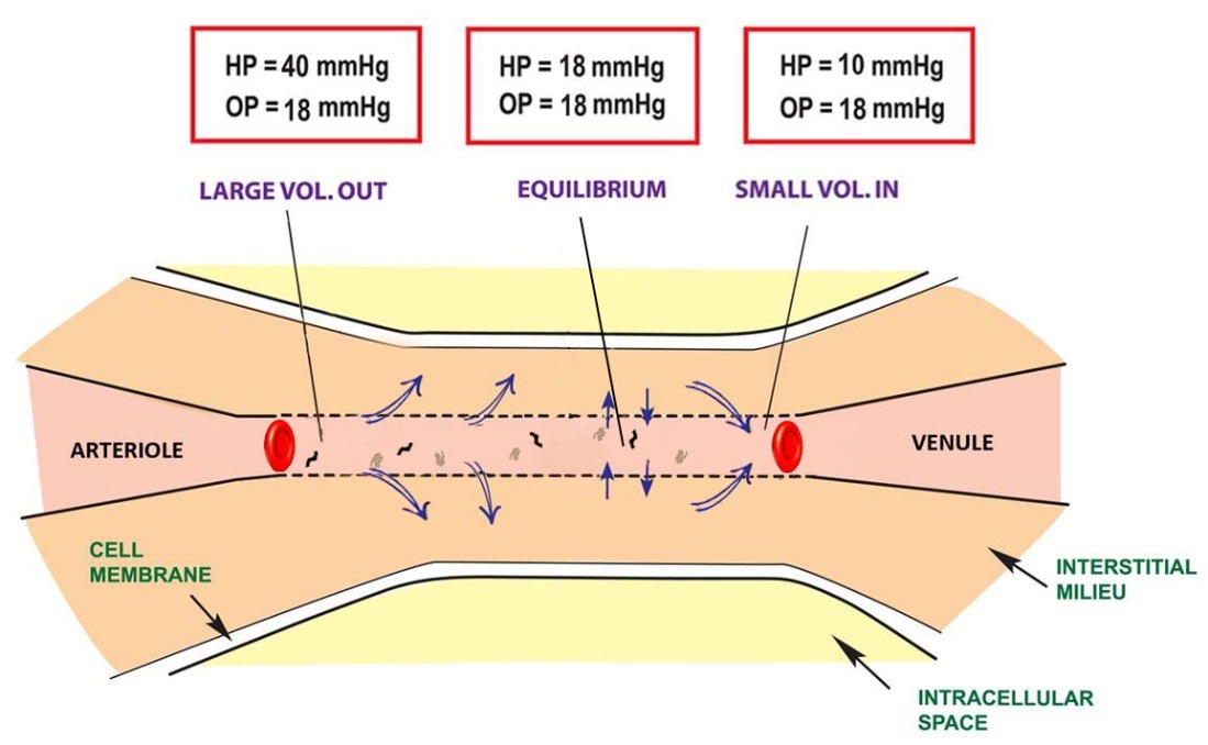
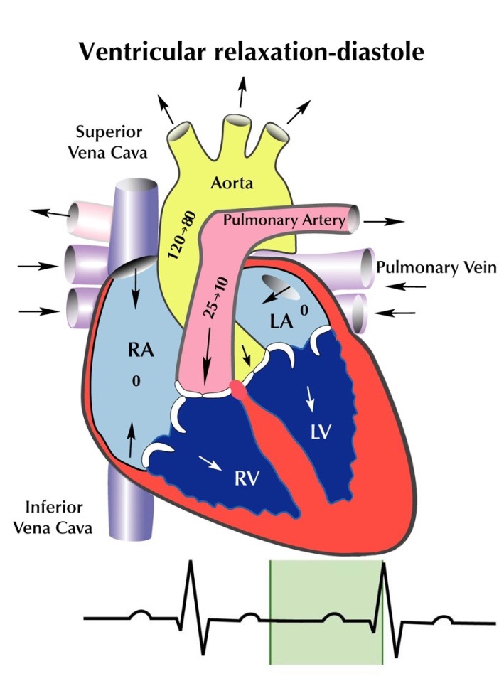
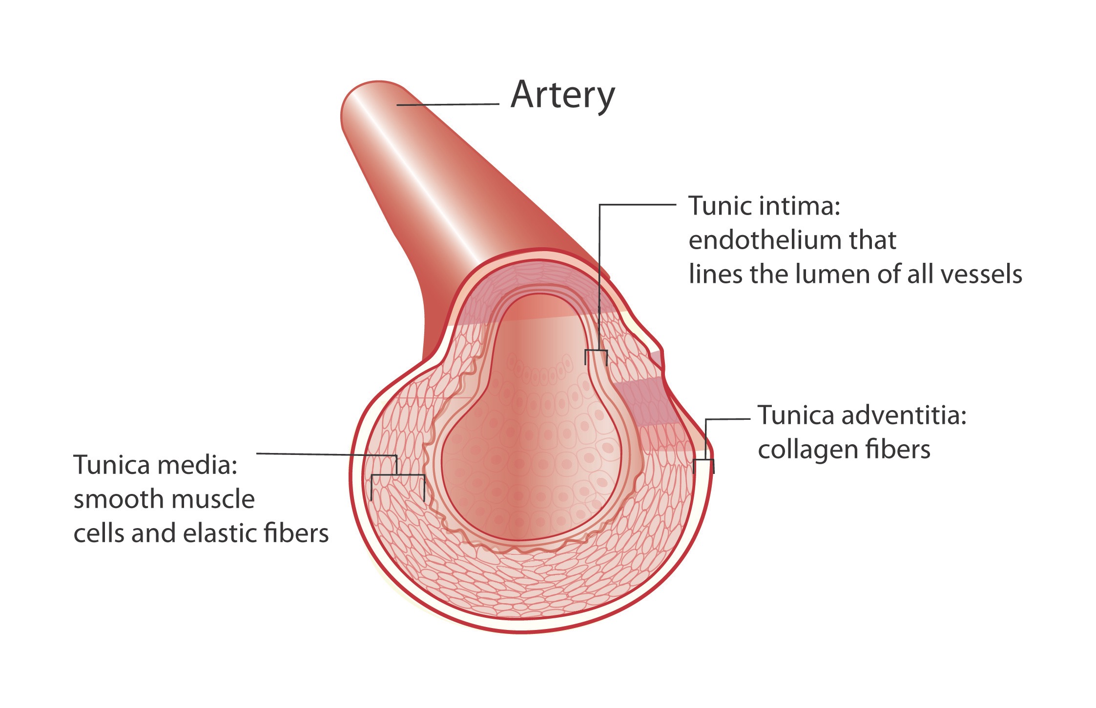
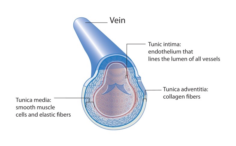
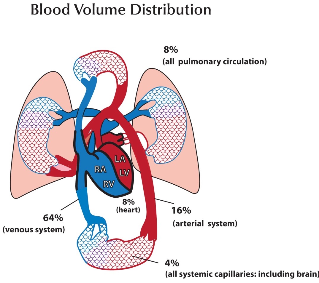
  There are three factors that determine blood flow to tissues: blood volume, “pump function” (cardiac function), and vascular capacitance and resistance.  Low blood volume, due to hemorrhage or dehydration, results in a decrease cardiac output because there is a decrease in stroke volume. When hydrostatic pressure is reduced to the point that perfusion to the body is compromised, we call this hypovolemic shock. Cardiogenic shock is poor tissue perfusion as a result of the heart not functioning well. This decrease in cardiac output can be the result of decreased stroke volume (e.g. from a heart attack and impaired contractility of the heart muscle), a low heart rate (e.g. from an unfavorable drug effect), or valve dysfunction, where forward flow of blood is impeded.  Vasogenic shock refers to decreased perfusion to the core of the body because of some problem with vascular tone. Under normal circumstances there is vascular tone, or slight vasoconstriction, to the extremities. This is because they do not need a lot of blood flow when at rest. When we exercise, additional flow is needed in the extremities and thus we increase our cardiac output, by increasing our heart rate, to accommodate this need. There are conditions, though, that occur when there is abnormal dilatation of the blood vessels (e.g. toxic effect of drugs), where perfusion to the core is compromised because of dilatation of arteries to the periphery. In the case of bad infections, sepsis, there can also be leakage of fluid from capillaries because of effects of certain conditions created. In this instance, intravascular proteins can be leaked into the interstitial space and fluid will be drawn out of the vascular space (since there is a loss of intravascular oncotic pressure). These vascular problems result in vasogenic shock.

There is one more type of shock that does not fit neatly into any of the above categories and that is something commonly referred to as mechanical shock. In this instance, there is impedance of blood flow as a result of something physically impairing blood flow. This can be the result of a collapse of the lung causing a tension pneumothorax, pulmonary emboli, cardiac tamponade, or some kind of mass or tumor compressing a major vessel from the outside and impairing flow. Again, when any of these conditions exist, we refer to this kind of shock as, mechanical shock. 

The human body has some ability to compensate for adverse conditions leading to shock, e.g. heart rate can increase to improve cardiac output, blood vessels to the extremities can constrict to shunt blood preferentially to important organs (heart, lungs, and brain), and the nervous system can give a sense of thirst and distress.  Compensatory mechanisms, however, can quickly become overwhelmed, and a state of non-compensatory shock sets in.  If conditions do not improve promptly, terminal shock ensues, organs fail, and the victim dies.  These are the 3 stages of shock: 1. Compensatory, 2. Non-compensatory, and 3. Terminal.  Compensatory shock can be subtle.  The patient may be mildly anxious, diaphoretic (sweaty), and tachycardic (defined as a heart rate greater than 100 beats per minute in a normal adult).  The blood pressure will be normal or near normal (systolic pressure about 110 to 130 mmHg for normal adult) and the patient will be mentating normally.  During compensatory shock, the body is releasing endogenous catecholamines (e.g. epinephrine from the adrenal glands) in an effort to increase cardiac output and constrict blood vessels leading to the skin and extremities and shunt blood preferentially to the abdomen, chest, and brain.  In addition, the kidneys are attempting to retain as much fluid as possible, so urine output decreases.  As conditions worsen, compensatory mechanisms begin to fail.  Catecholamine stores are depleted, renal function fails because of poor perfusion, and the patient’s blood pressure drops further.  If the cause of shock is not targeted soon and treated, the patient will progress to terminal shock and ultimately die.  Terminal shock is a condition in which cellular function is actively failing because of poor perfusion to the tissues.

The treatment of shock is straightforward: identify that the patient is in shock, find the cause of shock, and treat the underlying condition of causing shock.  After stabilization of the patient’s condition, provide supportive care to sustain a more normal physiologic state.  With this fund of new knowledge, let’s take a look at our original case scenario that started this chapter and see if we can save his life!

Capillary Physiology

The cells of the body require a continuous supply of oxygen and nutrients to survive.  They also require a process by which wastes can be carried away.  It is the role of the circulatory system to carry out these vital functions, and it is at the level of the capillaries that this process takes place.  Capillaries do not exchange directly with the intracellular compartment, but instead with an interstitial space that lies between the capillary and the cell membrane.


This interstitial space is a milieu of nutrients and wastes that are continually being exchanged by the intracellular and intravascular forces acting upon them.  Capillary circulation reaches every living cell in the body, via the interstitial fluid, and allows the exchange of nutrients and wastes.  Though there are different types of capillaries, which allow different amounts of exchange with the interstitial space, the general principles of diffusion remain the same and will be discussed in general terms.  Capillaries are thin walled and allow small particles and water to flow freely from the intravascular space to the interstitial space through tiny slits following diffusion gradients.  Large particles (e.g.  blood cells, proteins, and other large masses) are trapped within the vascular space.  These “trapped particles” create an osmotic gradient (oncotic pressure) that draws fluid back into the vascular space.  That is to say, oncotic pressure counteracts the hydrostatic pressure that drives fluid out of the vascular space.  There are four factors then, under normal circumstances, which determine the diffusion of water and other particles across a capillary wall:

  1. Particle Size
  2. Hydrostatic Pressures
  3. Oncotic Pressures
  4. Concentration Gradients

Under normal circumstances, fluid, electrolytes, oxygen and other nutrients flow out of the proximal portion of a length of capillary, and into the interstitial milieu that exists surrounding all cells (Figure 16).  As blood travels further through this length of capillary, hydrostatic pressure decreases to a point at which it equals oncotic pressure, and there is equilibrium of exchange to the point that only diffusion gradients affect the transport of particles across the capillary wall.  At the end, or distal portion, of a length of capillary, oncotic pressure is greater than hydrostatic pressure, and fluid is drawn back into the intravascular space (Figure 16).

EquilibriumBetweenIntravascularAndInterstitial-figure16

Figure 16
A normal state of equilibrium between the intravascular (filled with large particles, such as proteins, that creates an “oncotic” pressure) and interstitial spaces.

When hydrostatic pressures are high, there will be more fluid transported to the interstitial space (Figure 17).  When hydrostatic pressures are low, there will be more fluid transported into the intravascular space (Figure 18).  When oncotic pressures are low (e.g. in cases in which the liver does not produce enough albumin, such as in patients with liver failure), even with normal hydrostatic pressures, there will tend to be more fluid transferred to the interstitial space (Figure 19).  These principles are important because normal intracellular function depends on a very narrow range of fluid balance, electrolyte concentrations, and pH, and requires a continuous supply of oxygen and nutrients.  If any of these are disrupted, it will lead to cellular dysfunction and ultimately to cell death as conditions worsen.

EquilibriumBetweenIntravascularAndInterstitial-figure17

Figure 17
Elevated hydrostatic pressures in the capillary moves the point of equilibrium of Hydrostatic and oncotic pressures to the right (toward the venous end of the capillary) and allows less time for fluid and wastes to re-enter the circulatory
system. This leads to fluid retention in the tissues (edema).

EquilibriumBetweenIntravascularAndInterstitial-figure18

Figure 18
In a state of low hydrostatic pressure, the intravascular oncotic pressure is relatively higher thus the equilibrium between these opposing forces is moved to the left. This results in greater movement of fluid from the interstitial space to the intravascular space.

EquilibriumBetweenIntravascularAndInterstitial-figure19

Figure 19
When there are less intravascular osmotically active particles (less protein), this results in a state of low oncotic pressure. The hydrostatic and oncotic pressures equal each other closer to the venous end of the capillary. This results in a greater amount of time for fluid to escape from the vascular space and thus leads to tissue edema, much like high hydrostatic pressure does. Note: We don’t discuss a state of high oncotic pressure because it would be a very rare condition in which that would occur. If it did happen, though, what would the illustration look like?

Concentration gradients (Figure 20) exist between the interstitial space and the intracellular space because of complex “pump systems” that exist on the cell membrane of every human cell.  One of the most important pump systems is the Na+/K+ ATPase pump.  This pump creates an osmotic gradient between the high concentration of Na+ outside the cell, and the high concentration of K+ inside the cell.  It takes a lot of energy for cells to maintain these concentration gradients, and these gradients are necessary to optimize intracellular enzymatic function and to facilitate transport of certain interstitial particles into the cell.  There is an equilibrium between the intravascular space and the interstitial space such that the concentrations of most substances are essentially equal to each other, except for those particles that cannot escape the intravascular space because of their large size (e.g. blood cells, protein molecules).  We will discuss this topic again in the renal chapter.

ConcentrationGradients-figure20

Figure 20
The intravascular (plasma) space and the interstitial space share similar concentrations of ions, with sodium (Na+) being the primary cation and Chloride (Cl-) and bicarb (HCO3-) being the predominant anions (electrolytes measured in meq/L). The intracellular space has a much different
composition of ions because the cell membrane tightly controls the movement of charged particles into and out of the cell. The primary intracellular cation is potassium (K+) and the predominant intracellular anions are organic phosphates and proteins.

Exploring Medicine – A Foreword

I remember clearly the first meeting I had with Steve Jameson in summer 2005. I had just returned from a regional meeting of the National Association of Advisors in the Health Professions (NAAHP) where a number of sessions had focused on the importance of having health professions advisors build partnerships with health care practitioners in the community. So, when approached by Steve I was more than glad to meet to discuss what I thought was merely an opportunity to enhance our students’ access to shadowing opportunities.

Although that original meeting has led to much greater student access to clinical experiences, then I had no idea that it was the launching pad for an innovative educational experiment in the health sciences that has deeply contributed to the academic, professional, and personal development of hundreds of students – Exploring Medicine. In our first conversations in 2005, Steve proposed to teach a course at the College of Saint Benedict/Saint John’s University (CSB/SJU) that would show students how to critically think like a doctor and how to apply the material they were learning in basic science courses to the process of clinical diagnosis.

We eagerly supported Steve’s idea and in spring 2006 Exploring Medicine was taught for the first time. Since then it has been taught every spring at CSB/SJU and more recently in the fall semester at the University of St. Thomas. Exploring Medicine is truly a gem since it allows students to critically engage in a focused and structured learning experience from a clinical standpoint. Through Steve’s interactive lectures students develop analytical skills that allow them to experience the thinking process of a clinician making a diagnosis; through the panels of healthcare professionals and guest speakers students gain an appreciation for the diversity of the healthcare field and the necessity for a team-approach in care; and last but not least, through the structured shadowing experiences provided by the class students can see how principles covered in the course are directly applied in the process of diagnosis and patient care. Indeed so many students continue to pursue the relationships they have established with clinicians in Exploring Medicine that at CSB/SJU, with Steve Jameson’s help, we established a year-long internship program at the St. Cloud Hospital Emergency Department entitled the Student Health Assistant Program. Engaging community physicians to teach at local universities pays dividends, and Exploring Medicine is the ideal platform to establish those relationships.

The uniqueness of Exploring Medicine is that it is not static – Steve Jameson is continuously editing and tailoring his presentations, creating new experiences for students, and providing novel leaning tools and settings. Since 2006 Exploring Medicine also came to include this book – it is used for the class, but many Exploring Medicine alums will also vouch for its continued value as a refresher and review tool in their further studies. More recently Steve has developed online resources that allow Exploring Medicine to be delivered in its unique and creative fashion to health professions students in any college campus throughout the country; tools that were recognized by the AAMC with its 2013 iCollaborative award in biology.

In short, Exploring Medicine is much more than a course, a book, or a set of online tools. Exploring Medicine is a unique experience that allows health professions students to directly bridge their academic background to a structured clinical setting, and to begin to experience the intellectual world as seen through the eyes of a physician. In the process of helping Steve implement his vision at CSB/SJU, I have seen hundreds of students who are now working as physicians, physician assistants, physical therapists, among others engage in their first meaningful clinical discovery in that setting. Exploring Medicine is indeed the bridge from the world of the humanities, social and natural sciences of our college campuses to the experiential setting of clinical medicine and practice.

Manuel Campos, Ph. D.

Professor of Biology, Preprofessional Health Advisor College of Saint Benedict|Saint John’s University

Preface

Choosing a career is one of the most important decisions you will make in your life. While a career in health care, and in medicine in particular, can be incredibly exciting and rewarding, the journey to that end can be an enormous physical, emotional, and financial drain. The decision to go into this field must be an informed one.

Many are enamored with the medical field and with “being a doctor” long before they know much at all about the practice of medicine. Some are told, “You’re smart; you should be a doctor.” Others simply like what they see on TV. The Exploring Medicine series of modules will primarily focus on what it is like to be a physician, but the information is relevant to all that are seeking a career as a health care professional. To make an informed decision regarding a career in medicine, you should first explore the medical field by seeing what physicians do and learn to think like a physician.

This module, and others in this series, will allow you to do just that. Starting with the very first topic, you will plunge into the world of clinical practice. There you will find patients with a variety of medical problems (based on actual cases), many with life-threatening and life changing emergencies that you will need to work through and solve in order to save your patient’s life and make them well. By the time you have finished this module you will have learned to think and problem-solve like a doctor, and you will understand the process of diagnosing and treating disease.

The medical model is all about diagnosis and treatment of disease, and in order to understand that you have to understand pathophysiology. Pathophysiology is the study of disease. The pathophysiologic process of a disease is the series of events that take place and conditions that develop that result in a particular disease. In order to treat disease, the physician must understand something about the pathophysiologic process that led to the development of that disease. By going through the systems based modules you will gain insight into the thought process physicians use to diagnose and treat disease, and how they help patients to become well. Each system based module begins with a general overview followed by a discussion of the pathophysiology of a particular disorder: e.g. the Respiratory module reviews anatomy and physiology of the Respiratory System, then goes into a discussion regarding the cause of asthma and its treatment. Other modules, like Social Determinants of Health, focus on wellness as it relates to society, social factors, and public policy. Exploring Medicine modules were made for the students that have taken biology, chemistry, sociology, psychology, and other science courses, and wondered, “Why do I have to learn this stuff?” These modules apply the student’s science knowledge to the real world by making correlates to clinical medicine.

Books have been written on what will be small elements of the Exploring Medicine modules, so we obviously cannot comprehensively cover every topic. The authors have distilled a massive volume of material down to several manageable lessons. Despite this editing, the amount of subject matter may still seem intimidating at first, but that is not the intent of the authors. Do not get stressed or anxious. The focus of these modules will be on general principles and concepts. In order to transition from simple anatomy and physiology to treatment of disease, for example, we must streamline our approach in order for the reader to get some appreciation of what it is like to be a physician. The following module will take you, the medical enthusiast, through a very narrow slice of medical training: from medical school, to residency training, to clinical practice. Armed with your newly found knowledge, you will be able to make clinical decisions based on true-to- life patient scenarios and help your patients become well or stay well. If you find this process compelling, as the authors do, then you may seriously want to pursue the exciting field of medicine.

Introduction to the Chapters – Life and Death, Homeostasis and Equilibrium

Death is not the enemy but occasionally needs help with timing. – Peter Safar, M.D. The “Father” of CPR

What is life? That may seem like a metaphysical question, but it’s actually not. It is a very real question for those that provide medical care, but the answer can be surprisingly complicated. As a physician, physician assistant, nurse, or nurse practitioner, you will likely have many occasions where you will be dealing with patients as they look to cross that line from life to death. In the following chapters we will discuss a variety of pathophysiologic processes that will alter the normal function of the human body.

Before we delve into specific disease processes, though, let’s take a look at a more fundamental principle of human physiology and how a disruption of normal processes can lead to chaos, i.e. entropy, on a microscopic (actually molecular) level and to death on a macroscopic level.

The human body is a complex network of systems that operate in unity to keep us alive and active. In order to maintain normal function the body seeks homeostasis and defies equilibrium. Cellular reactions take place under very specific conditions: a narrow range of pH, the correct concentrations of particular ions, proper amount of substrates, etc. Homeostasis is the set of conditions that the body establishes and maintains in order to function properly. It takes energy to maintain this homeostasis. This energy has to be consumed in the form of chemical bonds contained in the food we eat, converted to a usable form, transported throughout the body to all of its cells, and metabolized into a form of energy the cells can use: ATP. The wastes of these reactions then have to be removed. It is a complex network of integrated systems that accomplishes all of these tasks.

In Exploring Medicine, we will look at four of the body’s integrally related physiologic systems (Cardiovascular, Respiratory, Renal and Integumentary) as examples of how the body maintains homeostasis. We also explore what can happen when these systems fail, and what we, as medical care providers, do to restore normal function and reverse the process of dying. All of the chapters have questions to answer along the way and challenging scenarios to solve at the end of each chapter. Answers to these questions and further elaboration of concepts are in the appendices near the end of the book. In the chapter on Evidence Based Medicine, we will discover how the medical literature helps to guide a physician’s clinical practice. As you strive to learn more about the field of medicine, remember the words of Hippocrates (the final line of the Hippocratic Oath), “May I always act so as to preserve the finest traditions of my calling and may I long experience the joy of healing those who seek my help.”

Cardiovascular System Unit Quiz

This is an open book test.  You can click here to open the lesson in a new tab/window to review the lesson as you take the quiz.  If you close the quiz you will have to start all over with a different set of questions. You need to achieve a score of 90% or greater in order to obtain the certificate of completion.

Please ensure you have enough time to complete the entire quiz at one time.

Cardiovascular Review Lessons

The following scenarios are designed to walk the future physician through a variety of real life situations that highlight various types of shock states.  Read the introduction closely and work through the scenario to ultimately diagnose and treat this patient’s condition (Answers follow each lesson).


Cardiovascular Physiology

There are three major areas of importance with regard to cardiovascular physiology: the physiology of the heart, the physiology of the large vessels (i.e. arteries and veins), and the physiology at the level of the capillaries.  As we discuss disease states (pathophysiology of the CV system), each of these subgroups of the CV system will have a unique role in the way it impacts the body.

Physiology of the heart:

Blood pulsates through the heart by way of organized contractions.  The ventricles relax after a contraction, the start of diastole, and the size of the chambers increase.  This action results in a dramatic decrease in pressure within the ventricular chambers.  The pulmonic and aortic valves (Figure 6a) then slam shut because there is a greater pressure in the pulmonary artery and aorta than there is in the right and left ventricles, respectively.  Blood then enters the ventricles from the atria through the tricuspid and mitral valves because there is greater pressure in the atria than there is in the ventricles.  The valves, under normal circumstances, prevent blood from going the wrong way during ventricular contractions, or systole.  During systole (Figure 6b), the ventricles contract and there is a rapid rise in pressure within these chambers.  The atrioventricular valves close, the pulmonary and aortic valves open, and blood is expelled into the pulmonary artery and aorta, respectively. Cardiac output is a term used to describe how much blood the heart puts out in one minute. Cardiac output (CO) is determined by the stroke volume (SV), the amount of blood ejected with each beat, and the heart rate (HR). The equation for cardiac output, then, is: CO = (SV) (HR), that is cardiac output equals stroke volume times the heart rate.

VentricularRelaxationDistole-figure6a

Figure 6a
Diastole is ventricular filling. Notice the ECG strip below the image. The shaded portion reveals the electrical activity of the heart in conjunction with the mechanical activity (ventricular relaxation) of the heart. We will discuss this concept in the following section.

VentricularContractionSystole-figure6b

Figure 6b
Systole is ventricular emptying as a result of contraction of the ventricles.


Atrial contraction is induced by propagation of a signal that begins in the sinoatrial (SA) node (Figure 7).  Electrical depolarization of the atrial myocardium stimulates an organized mechanical contraction of the muscle fibers and a resulting atrial contraction.  Under normal circumstances, the signal cannot travel directly from atrial myocardium to ventricular myocardium.  The signal must first pass through the atrioventricular (AV) node (Figure 7) where it is held for a fraction of a second to allow mechanical contraction of the atria to complete before contraction of the ventricle commences.  When the electrical signal is allowed to propagate from the AV node, it rapidly depolarizes the ventricular myocardium.  Ventricular contraction follows this depolarization.  As the ventricle contracts, the chamber pressure overcomes the pressure within the atria, and the atrioventricular valves close (the “lub” in lub-dub of the heart sounds).  As the ventricle continues to contract there is ventricular emptying, and the completion of systole.  With the initiation of diastole, the pulmonic and aortic valves close, and there is the characteristic “dub” heard on auscultation with a stethoscope. 

ElectricalDepolarization-figure7

Figure 7
Electrical activity in the heart is measured by electrodes on the skin and results in a characteristic electrical pattern on an ECG. Notice that the major spike in amplitude of this electrical signal is within the ventricles because there is much more heart muscle there conducting electricity than there is in the atria.

Depolarization of atrial and ventricular myocardium results in a characteristic electrical signal on an ECG (electrocardiogram, also known as an “EKG”).  Electrical sensors or “leads” are placed at various locations on the chest and extremities and the electrical signal propagating through the heart is read.  If all of the muscle tissue in the heart is healthy and getting an appropriate supply of oxygen and nutrients, it will conduct normally and give a normal electrical signal.  Physicians can determine, by reading a multilead (usually a “12 lead”) ECG (Figure 8), not only whether heart muscle is functioning properly, but also where any poorly functioning muscle is in the heart.

Normal12LeadECG-figure8

Figure 8
Normal “12 Lead” ECG

At rest, cardiac myofibrils have a negative electrical potential on the surface of the cell membrane of about -90mV.  When a proper electrical stimulus takes place, the entire surface of the cell membrane depolarizes (Figure 9a) then propagates the stimulus to adjacent myocytes until all of the heart muscle depolarizes in an organized fashion (Figure 9b).  This wave of depolarization can be detected by a skin electrode of an electrocardiogram lead (Figure 10).  When the majority of heart muscle cells are depolarizing in a direction toward a lead, it is picked up as a positive deflection on the electrocardiogram (ECG).  When the major wave of depolarization is away from an electrode, the ECG tracing reveals a negative deflection.  Leads are placed at strategic locations across the chest and extremities to give multiple “views” of the heart.  This allows the physician to determine if there is normal conduction of an electrical stimulus throughout the heart. 

Depolarization-figure9a

Figure 9a
Depolarization propagates along the length of the myofibril. Contraction subsequently ensues.

Depolarization-figure9b

Figure 9b
Propagation of the depolarization across  the atrial myocardium leads to contraction of the atria.

Depolarization-figure10

Figure 10
Depolarization toward a skin electrode is recorded as an upstroke on the EKG monitor (Figures 9a, 9b, and 10 reprinted with permission from Dale Dubin, M.D. In: Rapid Interpretation of EKG’s, 6th Ed. Fort Myers: Cover, 2000.)

The deflections produced on the ECG by depolarizing myocardium are given letter names for the “waves” that they produce (Figure 11).  A “P wave” results from depolarization of the atrial muscle.  The “QRS complex” is the name given to the deflections produced by depolarization of the ventricles.  The “T wave” is the electronic signal produced by the repolarization of the ventricles.  One does not see a repolarization wave for the atria because it is exceedingly small (flat) and typically buried in the QRS complex.

NamesofWavesonECG-figure11

Names of the waves on an ECG

When myocardium is not getting enough blood flow or is somehow injured, it does not conduct electrical signals normally. Ischemia, or decreased blood flow, to a portion of the heart is usually the result of an occlusion of the coronary artery serving that area of myocardium: typically a blood clot or “thrombus” (Figure 12).  This decreased blood flow can result in infarction (cell death) of the affected heart muscle in this region (Figure 13).  An acute myocardial infarction (“heart attack”) produces characteristic “ST elevations” on an EKG early in this event (Figure 14).  Depending on the location on this ST elevation on an EKG (Figures 15a and 15b), the physician can determine where the decrease blood flow to the heart is, and, typically, what coronary vessel is involved.  Knowing this allows the interventional cardiologist to quickly pass a long thin catheter to the site of the obstruction and open the occluded vessel (coronary angioplasty and stenting).

Infarction-figure12

Figure 12
A blood clot plugging a major artery serving heart muscle.
This leads to a heart attack or “myocardial infarction.”

BloodClotIllustration-figure13

Figure 13
An illustration showing the effect of a blood clot in a
major coronary artery. (Figures 12 and 13 reprinted with permission from Dale Dubin, M.D. In: Rapid Interpretation of EKG’s, 6th Ed. Fort Myers: Cover, 2000.)

STElevation-figure14

Figure 14
ST elevation: indicative of heart attack on an ECG
(Reprinted with permission from Dale Dubin, M.D. In:
Rapid Interpretation of EKG’s, 6th Ed. Fort Myers: Cover, 2000.)

12LeadECGAcuteAnteriorMyocardialInfarction-figure15a

Figure 15a
12 lead ECG indicating an acute anterior myocardial infarction. Notice the ST segment elevation in leads V1,V2, & V3, and even extending all the way to V6. This finding indicates to the physician that there is a vessel in the anterior (front) portion of the heart that is plugged.

12LeadECGAcuteInferiorMyocardialInfarction-figure15a

Figure 15b
12 lead ECG indicating an acute inferior myocardial infarction. Notice specifically the ST segment
elevation in leads II, III, and AVF, among changes elsewhere. This indicates that there is an occlusion of a coronary artery serving the inferior (lower) portion of the heart.

Physiology of the large vessels (arteries and veins):

Arteries and veins are not mere conduits through which blood travels, but are part of a complex system that regulates blood flow to various tissues and helps regulate blood pressure.  In fact, most drugs used to treat hypertension and shock are directed at influencing the action of these vessels (primarily the arteries).  In certain tissues, e.g. skin and various appendages (fingers, toes, ears), tiny arteriovenous anastomoses can shunt blood past specific capillary beds.  This action can preserve circulation to vital organs and help maintain body heat when needed.  The reverse is also true when the body needs to dissipate excess heat (can shunt more blood to skin by opening flow to the microcirculation of the skin).  Smooth muscle in the wall of the artery allows the vessel to change its caliber.  When these muscles contract, there is a rise in arterial blood pressure proximal to the constricting vessels, and when these vascular smooth muscles relax there is decreased blood pressure proximal to the dilating vessels.  Blood pulsates through the arteries as a result of ventricular contraction.  There is a rapid rise in intravascular pressure as blood is ejected from the heart, and a gradual decline in blood pressure as blood is distributed to capillary beds.  The peak pressure in the artery is called the systolic pressure and the lowest pressure in the artery is called the diastolic pressure. 


Blood pressure is a function of flow and resistance. If either flow or resistance goes up, blood pressure goes up, and vice versa. Catecholamines, such as epinephrine and norepinephrine, can be produced and secreted into the blood stream from the adrenal glands (endogenous supply) or injected into an intravenous (IV) line (exogenous supply).  These catecholamines can have a profound effect on blood pressure and pulse because of the unique characteristics of these agents.  They can cause the heart to beat faster and stronger and blood vessels to constrict.  When arteries constrict, they reduce blood flow to the tissues distal to the constriction, but increase the blood pressure in the central circulation, preserving blood flow to major organs such as the heart, lungs, and brain.  Other tissues can sustain a decrease in blood flow for a longer period of time than these vital structures, however, sustained poor tissue perfusion, i.e. shock, leads to cell dysfunction, and in severe uncorrected cases, death of the cells and even the patient.

Cardiovascular Anatomy

The CV system is a complex network consisting of the heart, arteries, veins, capillaries, and blood.  The heart is probably the first thing that comes to mind when you think of circulation; it is, after all, the pump that delivers blood throughout the body.  Arteries and veins carry blood that travels away from and back to the heart, but they are not simply conduits through which blood flows.  These vessels have a smooth muscle layer that allows them to dilate and constrict depending on the immediate physiologic needs of the various tissues.  These large vessels and the heart together determine the hydrostatic pressure within the capillary vessels.  It is at the capillary level that exchange of fluid and solutes between the blood and the interstitial space takes place.  The interstitial fluid then exchanges its contents directly with the cells of the body.  This fulfills the purpose of the circulatory system: to bring nutrients to and wastes from the cells and tissues of the body.  One would conclude, then, that the real work of the CV system is done at the level of the capillaries.  In order for the work to get done at the level of the capillaries, all of the components of the CV system have to be functioning properly.  We will go through the normal anatomy and physiology first then discuss what can go wrong: i.e. discuss pathophysiology.

Anatomy:

The human CV system (fig. 1) is composed of a heart, about 60,000 to 100,000 miles of blood vessels, and about 6 liters of blood (in the average adult).  The blood vessels consist of thick walled, high pressure arteries and arterioles, thin walled, low pressure veins and venules, and microscopic, very thin walled capillaries.  This is a complex system that continually interacts with every part of the body.

HumanCVSystem-figure1

Figure 1
The major vessels of the human circulatory system

The human heart (Figures 2a and 2b) is a four chamber pump that moves blood through two separate circulatory systems within the body: the pulmonary circulation and the systemic circulation.  The left and right atria are two low pressure chambers at the upper portion (base) of the heart that collect venous blood.  At the lower portion of the heart are two high pressure, thick walled chambers: the ventricles.  The heart can be divided down its center into a right and left side, each containing one atrium and one ventricle.  These two “sides” are referred to as pulmonary circulation (right side) and systemic circulation (left side).  Valves between the atria and ventricles (the atrioventricular valves) allow blood flow from the atria into the ventricles, but not the reverse.  Valves between the ventricles and the pulmonary and systemic circulation (the pulmonary valve and aortic valve, respectively) allow blood to flow into these outflow arteries, but not the reverse.  Pulmonary circulation is a lower pressure system than the systemic circulation, thus the muscular wall of the right ventricle is much thinner than that of the left ventricle.  The aorta is the largest artery in the body and it carries blood to the systemic circulation.  The first arterial branches off of the aorta are the coronary arteries.  These vessels provide blood flow for the delivery of oxygen and other nutrients to the heart tissue.  About 8% of the total circulating blood volume is in the heart at any given time (Figure 4).

ExternalViewofHeart-figure2a

Figure 2a
External view of the heart. The blood vessels
on the outside of the heart are the coronary arteries and veins.

InternalViewofHeart-figure2b

Figure 2b
Internal view of the heart. The atrioventricular (AV) valve between the RA and RV is the tricuspid valve, and the AV valve between the LA and LV is the mitral valve. In this depiction the pulmonary and aortic valves are open, so there is blood flow across them. The arrows indicate the path of blood flow through the heart.

Arteries (Figure 3a) are defined as vessels that carry blood away from the heart.  These are high pressure vessels that are, out of necessity, thick walled.  Much of the thickness of the arterial wall is due to smooth muscle.  Many conditions, hormones, and drugs can make the caliber of the artery change depending on whether the muscles are stimulated to contract (and decrease the caliber of the vessel) or relax (and increase the caliber of the vessel).  All blood vessels (and the heart too) are lined with a thin endothelium.  This is a very important barrier to the underlying vessel tissue and it plays a major role in keeping blood flowing smoothly through the vessel.  About 16% of the blood in the body is circulating through the arterial vessels at any point in time (Figure 4).

Artery-figure3a

Figure 3a
Microanatomy of an artery

Vein-figure3b

Figure 3b
Microanatomy of a vein (notice the thinner muscular layer and the presence of valves)

BloodVolumeDistribution-figure4

Figure 4
Notice that despite the fact that capillaries make up the vast majority of the over 60,000 miles of blood vessels in the body, they only hold 4% of all of the blood. The veins hold the vast majority of the body’s blood.

Veins (Figure 3b) are defined as vessels that carry blood to the heart.  This is a high capacity – low pressure system, and thus these vessels have thin walls.  Veins have little muscle in their walls, so they have little ability to change their caliber and thus the capacity of this space.  We usually think of veins as the blue vessels (but the blood is not actually blue) on diagrams because they carry deoxygenated blood from the systemic circulation back to the heart: specifically, the right side of the heart.  The exception to this rule is blood returning to the left side of the heart via the pulmonary veins; this blood is richly oxygenated and typically depicted in red (and the blood is actually bright red in color).  The majority of blood in the body, about 64 % (Figure 4), is contained in veins (a high capacitance system).

Capillary circulation makes up the vast majority of the thousands of miles of blood vessels in the body.  Despite that fact, only about 4% of total body blood volume is contained in the capillaries (Figure 4).  Capillaries (Figure 5) are one cell layer thick, being made up of only an endothelial layer and a basement membrane.  They are narrow tubes, measuring only 7 – 10 microns in diameter.    A red blood cell, for comparison, is about 8 microns in diameter (and about 2.4 microns thick).  Red blood cells often have to deform somewhat simply to get through some capillary beds.

Capillaries-figure5

Figure 5
Particles small enough to get through the cleft can freely exchange with the interstitial space. Larger particles are trapped within the capillary.

Blood is made up of cells and serum, and we will keep it that simple.  The cellular components of blood are red blood cells, white blood cells, and platelets.  Within red blood cells is hemoglobin, a complex protein that is capable of carrying gasses such as oxygen and carbon dioxide.  Serum has a whole host of elements, but we will focus on those that affect osmotic activity (e.g. electrolytes and protein) since these elements will affect exchange of fluid and substrates at the level of the capillaries.

Introduction to CV & Patient Scenario

Cardiovascular System

At a cardiac arrest, the first procedure is to take your own pulse.*

Author, Samuel Shem, M.D.

Introduction:

There is no more exciting topic in medicine than the cardiovascular (CV) system: that is the reason why it leads off as the first topic in Exploring Medicine.  Life starts and stops, literally, with this system.  When you are the physician charged with the responsibility of caring for a critically ill patient, you must have a strong working knowledge of this system.  In order to cover a topic as broad as this in a single chapter, an emphasis will be placed on just the key elements as we break this system into a few basic parts.  The goal of this chapter is to have you develop enough of an understanding of the fundamentals of the CV system that you will be prepared to discuss the designated pathophysiologic** process at the end of the chapter: shock. 


You are the emergency physician on duty in a hospital Northern Minnesota.  It’s been a relatively quiet night until suddenly, through the ambulance bay doors, comes a woman screaming for help.  You, and the two nurses working with you, run to the car that the woman drove to the hospital.  You find a middle aged, pale looking male slumped against the front passenger door of a car.  He is diaphoretic (sweaty), marginally responsive as you try to talk to him, and clearly struggling to breathe.  You quickly move him to a gurney and wheel him to your resuscitation room.  What do you do?

Jot down some notes here as to what you think you would do as the emergency physician in this case, and then compare this to what you would do after reading this chapter.

Natural Science Module CV

Cardiovascular System

At a cardiac arrest, the first procedure is to take your own pulse.*

Author, Samuel Shem, M.D.

Introduction:

There is no more exciting topic in medicine than the cardiovascular (CV) system: that is the reason why it leads off as the first topic in Exploring Medicine.  Life starts and stops, literally, with this system.  When you are the physician charged with the responsibility of caring for a critically ill patient, you must have a strong working knowledge of this system.  In order to cover a topic as broad as this in a single chapter, an emphasis will be placed on just the key elements as we break this system into a few basic parts.  The goal of this chapter is to have you develop enough of an understanding of the fundamentals of the CV system that you will be prepared to discuss the designated pathophysiologic** process at the end of the chapter: shock.